Lung Cancer Mystery in Non-Smokers

One of the emerging mysteries around lung cancer is why it occurs so often in people who don’t smoke – particularly in Asia. A new paper in JAMA Network Open investigates the phenomenon by presenting results from South Korea indicating that established screening criteria would have missed nearly two-thirds of lung cancer patients.

From its earliest days, CT lung screening has been targeted at heavy smokers who have the greatest chance of developing lung cancer.

  • Risk-based criteria determine who should be screened, usually based on some metric of smoking history (such as the USPSTF’s 20-pack-year threshold).

But many countries that have had national lung screening programs in place for a while – mostly in Asia – are learning that lung cancer also occurs in people who have never smoked at all. 

  • Whether and how to screen these individuals has become the subject of debate. 

In the current study, researchers from South Korea examined 89.9k patients who were diagnosed with lung cancer from 2013 to 2018, just before the country started its national lung screening program in 2019. 

  • Researchers focused on whether patients would have been eligible for screening under established criteria, such as from the USPSTF, ACS, and NCCN.

Among people diagnosed with lung cancer…

  • 65% would not have been eligible for lung screening under international guidelines. 
  • 44% had no smoking history. 
  • Screening eligibility differed sharply by sex, with men eligible at far higher rates than women (57% vs. 2.3%).
  • Smokers who weren’t eligible for screening had 5% lower rates of all-cause mortality and 4% lower lung cancer-specific mortality than smokers who were.
  • People who had never smoked had 14% lower rates of both all-cause and lung cancer-specific mortality. 

The data point out an interesting dilemma inherent in CT lung cancer screening, which from the outset was set up to be risk-based rather than population-based like mammography to avoid many of screening’s downsides, like overdiagnosis.

  • But that approach is missing many people who would otherwise benefit from screening, especially as we learn more about the elevated risk among many non-smoking populations, such as East Asian women.

The Takeaway

The new study shows momentum building toward broader CT lung cancer screening criteria that go beyond smoking status. The question is how quickly such criteria are adopted, and whether they gain traction outside of Asia, where recognition of lung cancer among non-smokers is growing.

Your Reporting Platform Is Now Your AI Strategy

By Sheela Agarwal, MD, MBA, a practicing radiologist and chief medical information officer at Microsoft 

For most of my career, the radiology reporting platform was a workflow tool. Today, it is an AI strategy, and the way we evaluate it must change with it. 

I see that from two sides. As a practicing radiologist, I live in the reporting workflow. As CMIO at Microsoft, I help shape how technology serves it. 

  • The platform underlying radiology reporting now determines something bigger than features. It determines how effectively AI is integrated into the workflow and where it is making the biggest impact, for both radiologists and the care patients receive.  

The pressure is real, and it is human. Imaging volumes keep climbing while the workforce to read images struggles to keep pace, and the cognitive load on every radiologist grows with it. 

  • The question is no longer whether to adopt AI. It is which foundation can carry AI reliably, study after study. 

Most reporting solutions were not built to be that foundation. PowerScribe One is. The proof is in the scale: 

  • 280+ organizations, proven across IDNs, academic centers, community hospitals, and independent practices. 
  • 10,000+ radiologists have made PowerScribe One part of how they work. 
  • 10M+ reports every month, a volume that reflects real, sustained use. 

That scale is not the point on its own. It is evidence that the technology delivers where it counts. What matters to a radiologist is more specific: does it lighten a heavy worklist, or just add another click? 

As an AI companion to PowerScribe One, Dragon Copilot builds on that foundation, bringing prior reports, patient context, and information from credible sources to the same screen as they read, so radiologists work with a more complete picture without leaving their workflow. 

  • The deeper value is what it frees radiologists to do: practice at the top of their license, spending less time on the mechanics of report creation and more on the complex reads and the diagnostic judgment that shape a patient’s care. 

A reporting platform must be ready for what comes next, evolving alongside radiology and AI. 

  • For organizations still on PowerScribe 360, now is the time to evaluate the path forward. Migration to PowerScribe One preserves existing workflows and configurations, allowing organizations to modernize at their own pace without disrupting care. 

The Takeaway 

This is not simply a radiology reporting system upgrade. It is choosing a partner committed to practical innovation, built on decades of workflow expertise and deep collaboration with customers and partners. Connect with Microsoft’s team or click here to learn more about PowerScribe One and Dragon Copilot.

Ultrasound and DBT Screening — Time to Call It Quits?

Ultrasound long ago carved out a role in breast cancer screening as a complementary tool to conventional 2D mammography. But is ultrasound still needed, now that U.S. breast screening programs have largely switched over to 3D digital breast tomosynthesis (DBT)? A new study in Academic Radiology raises questions. 

Conventional 2D mammography has well-known shortcomings, particularly in women with dense breast tissue that can obscure lesions. So alternative modalities like ultrasound, breast MRI, and contrast-enhanced mammography (CEM) are called in to help when needed.

  • Past research has shown that supplemental ultrasound can improve the cancer detection rate (CDR) in a 2D mammography screening program by 3-4 cancers per 1k women. 

But those studies were performed before the switch to DBT, which can often see around overlapping structures thanks to a gantry head that acquires multiple images as it pans across the breast. 

  • So is ultrasound still needed in screening programs using DBT? Researchers from Weill Cornell Medicine at New York-Presbyterian Hospital tested the hypothesis by examining 103k screening exams from 2014 to 2024 in which both DBT and ultrasound were used. 

In the study, researchers found that screening ultrasound after a negative DBT result…

  • Generated 1.9k biopsies, or 19 biopsies for every cancer the modality detected.
  • Produced an additional cancer detection rate of 1.0 per 1k women, compared to an additional CDR of 3-4 cancers for ultrasound in the 2D mammography era. 
  • Had an overall false-positive screening rate of 98.5%, a false-positive biopsy rate of 94.8%, and a positive predictive value of biopsies performed (PPV3) of just 5.2%.
  • Generated $652k false-positive screening ultrasound costs and $1.16M in ultrasound-guided biopsy costs.

The numbers are sobering and indicate that the days of ultrasound as a supplemental screening modality to DBT screening could be coming to a close. 

  • Instead, the researchers recommended that ultrasound screening be replaced by more sensitive modalities like breast MRI or CEM, both of which are fortunately more available now than during the 2D mammography era.

The Takeaway

The new study answers the question – in the negative – of whether supplementary ultrasound is still needed in the era of DBT screening. The positive subtext here is that the research confirms the improved detection performance of 3D compared to 2D mammography.

MRI for Prostate Cancer Staging

Digital rectal exams for prostate cancer staging could become a thing of the past thanks to MRI. A new study in JAMA Network Open found that MRI performed as well as digital exams in determining the extent of prostate cancer disease.

Prostate cancer screening is moving closer to becoming a more widely accepted test, and MRI has played a major role in that evolution by enabling more precise workup of men with high PSA levels.

  • But what about other aspects of prostate cancer diagnosis and treatment, such as staging men found to have clinically significant disease? It turns out MRI has a role to play there as well. 

To learn more, researchers from Germany looked at data from 4.4k men with a median age of 66 and median PSA level of 7.4 ng/mL (anything over 3 ng/mL is typically referred for additional workup). All patients were scheduled for radical prostatectomy. 

  • Patients received digital rectal exams to assign clinical T stage and assess characteristics of cancer severity, such as local tumor extent, extracapsular extension, and seminal vesicle invasion. 

This was compared to multiparametric MRI scans on 1.5T or 3T systems, with data reported using the PI-RADS scale. The study’s primary outcome was distant metastasis-free survival. 

Among the patient cohort, researchers found…

  • MRI staging was slightly more accurate than digital exams for predicting biochemical recurrence-free survival (C index = 0.62, with 0.5 representing chance-level prediction and 1 indicating perfect prediction). 
  • And MRI was also better at predicting distant metastasis-free survival (C index = 0.67).
  • But MRI and digital exams were comparable when using four of the major prostate cancer risk classification systems in Europe.

What to make of the results? MRI didn’t have a huge advantage over digital rectal exams, but it was good enough for the authors to suggest that digital exams could be eliminated in favor of MRI workup instead.

  • That would enable clinicians to avoid many of the digital exam’s shortcomings, such as subjectivity, operator dependency, and restricted ability to assess extracapsular extension or seminal vesicle invasion.

The Takeaway

This week’s study shows that MRI’s role in prostate cancer diagnosis and treatment goes beyond just screening, and the digital rectal exam’s role for patient staging could soon become a thing of the past in all but a few cases. 

Where Do Humans Belong in the AI Loop?

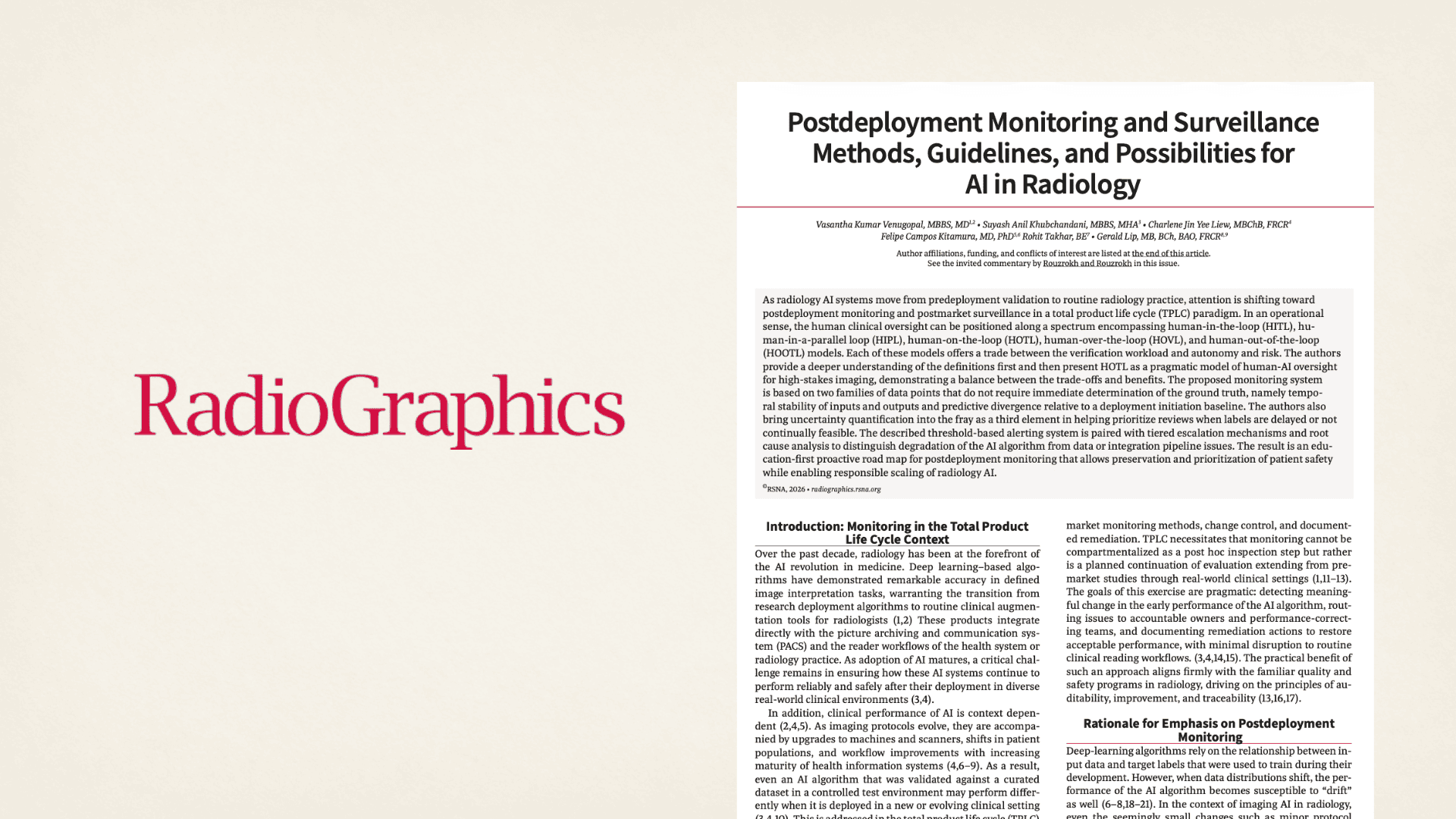
AI has its skeptics and superfans when it comes to interpreting medical images, inspiring a wide range of approaches to checking its work, but a new RadioGraphics paper argues the right level of oversight lies somewhere in the middle. 

There’s a strong consensus on the importance of monitoring AI post-deployment, even as the right level of rigor remains an open question. 

  • The FDA and many of its global peers require institutions to track AI performance, implement human oversight, and have a corrective action plan. 
  • Past studies show oversight mechanisms are top of mind for many radiologists. 

The international researchers behind the article say a human-on-the-loop (HOTL) model is the sweet spot for radiology departments as they balance safety and reliability with efficiency. 

  • Under this framework, radiologists don’t have to review every AI output.
  • At the same time, the proposed model doesn’t take AI results at face value.
  • HOTL threads the needle by alerting humans to drift and accuracy problems.
  • If performance drops, the work of reading these scans reverts to radiologists. 

The model acknowledges that even the most cleverly designed radiology algorithms can be sensitive to changing inputs.

  • Scanner upgrades, workflow adjustments, and shifts in patient mix can leave AI that aced training tests out of step with clinical realities. 

HOTL has its perks, but the authors note a few challenges. 

  • Excessive alerts risk desensitizing monitoring teams, so it’s important not to set the notification threshold too low or treat minor deviations as urgent. 
  • Declines in subgroup performance can go unnoticed if overall metrics stay stable.

The Takeaway

For radiology departments, the ideal AI oversight plan protects patients without making the technology more trouble than it’s worth. It seems the HOTL system outlined in RadioGraphics checks those boxes by focusing on trends over individual outputs. Even so, the debate over how to best supervise AI is sure to remain lively. 

Top 6 Radiology Trends for the First Half of 2026

The first half of 2026 is now in our rear-view mirror. As we do every year, we’ve compiled a list of the top six stories – one for each month – to help recap what was important in medical imaging.

Radiology Reporting Booms as Microsoft Sunsets PowerScribe 360
Microsoft’s announcement in February that it would be sunsetting its PowerScribe 360 radiology reporting software set off a scramble for market share that continues months later. While PowerScribe was instrumental in moving radiology to speech recognition-based reporting, many radiology facilities are seeing the announcement as a chance to adopt more modern AI-powered reporting solutions.   

Radiology Dominates List of New AI Approvals
The FDA regularly updates its list of AI-enabled medical devices with marketing authorizations, and our coverage of the agency’s decisions was the second most-popular story of 2026’s first half. As with previous updates, radiology dominated the list, garnering 76% of all authorizations since the agency began keeping count and 75% in the fourth quarter of 2025.

Residency Push Skips Radiology
Workforce shortages are a hot story across healthcare, and radiology is no exception. But the specialty won’t be getting much help from a federal initiative to add more resident training slots. Of the more than 400 residency programs awarded funding so far, only two diagnostic radiology programs were selected.

Radiologist Quit Rates Double in a Decade
Having to do more work with less personnel could be convincing many radiologists to leave the profession. Our readers paid close attention to a February story on a JACR study that documented a doubling of the radiologist quit rate over 10 years, and the exact point in terms of case workload when rads were most likely to leave. 

Data Is Lacking on AI’s ROI
As radiology AI slowly moves from pilot projects to widespread clinical adoption, a new survey reveals a paradox: The technology is popular with radiologists, but few imaging facilities using AI have collected hard data showing its return on investment. That’s according to another popular story from April

Study Finds Variation in Radiologist Workload
Our sixth and final top story of 2026 addressed the growth in imaging volume since the COVID-19 pandemic, and how radiologists responded. Researchers found that volume did indeed grow faster than the supply of radiologists, but some imagers were doing more than others in picking up the slack. 

The Takeaway 

Our readers have apparently been interested in workload issues so far this year, as evidenced by the fact that three of the top six stories on The Imaging Wire for the first half of 2026 had something to do with radiology’s rising exam volume and its ramifications. The other half broadly addressed AI and imaging IT issues. See a connection?

Mammo Modality Face-Off for Early Breast Cancer

When it comes to early breast cancer detection, which medical imaging modality is best: full-field digital mammography, digital breast tomosynthesis, or breast MRI? A new study in Clinical Radiology picks winners – and brings the receipts. 

Breast imagers are fortunate to have many technologies at their disposal, each with its own strengths and weaknesses. 

  • X-ray-based mammography tools like FFDM and DBT are easily available and relatively low cost, while breast MRI delivers the highest resolution but is expensive, less available, and more time-intensive to perform. 

So when does it make sense to use each modality? Researchers from China tested four techniques – FFDM, DBT, and breast MRI at 1.5T with accelerated and full protocols – in 329 patients with early-stage breast cancer (maximum tumor diameter ≤ 2 cm). 

  • They also analyzed results according to breast tissue density, as dense breast tissue is not only a cancer risk factor but can also obscure lesions on X-ray-based modalities.

Across the study sample, researchers found…

  • There was little difference in sensitivity between the four techniques for women with non-dense breast tissue, with FFDM, DBT, and accelerated breast MRI achieving 91% compared to 94% for full-protocol breast MRI.
  • But breast MRI pulled ahead in sensitivity for women with dense breast tissue, both with accelerated and full protocols (95% and 94%) beating DBT and FFDM (90% and 83%).
  • Accelerated breast MRI had performance comparable to the full protocol regardless of breast density, but at almost half the median scan time (8 vs. 15 minutes).
  • Accelerated and full-protocol breast MRI had the same specificity (94%), ahead of both DBT and FFDM (88% and 83%).

What to make of the results? Researchers said the findings in women with non-dense breast tissue reinforce that X-ray-based modalities are sufficient.

  • For women with dense breast tissue, accelerated breast MRI offers performance close enough to the full protocol that breast imaging practices can feel comfortable offering the faster exam.

The Takeaway

It’s no surprise that breast MRI beat both FFDM and DBT mammography for early breast cancer detection in women with dense breast tissue. But it is intriguing that there wasn’t much difference between breast MRI with either accelerated or full protocols. That’s good news for practices that want to make this powerful modality accessible to more women. 

Get every issue of The Imaging Wire, delivered right to your inbox.