Could MRI Reduce Prostate Biopsies?

Could MRI one day reduce the need to biopsy some patients with suspected prostate cancer before they get treatment? U.K. researchers raise that intriguing prospect in a new study in European Journal of Radiology

Biopsy is an integral part of the workup protocol for men suspected of having prostate cancer, typically due to their elevated PSA levels.

  • But prostate biopsy is an invasive procedure, and can cause both complications and anxiety in patients, as well as contribute to higher healthcare costs.

MRI has proven effective in selecting which patients should be sent for biopsy and which ones can be followed with active surveillance – thus avoiding biopsy’s negative consequences.

  • But what if you could eliminate biopsy altogether in some men by making the diagnosis directly with MRI, and sending patients straight to treatment? 

That’s what researchers from University College Hospital in London tried in the new EJR paper by studying 880 consecutive patients who presented to their prostate clinic and got multiparametric MRI scans. Of these, 251 were considered suspicious for disease based on their scans.

  • Radiologists interpreted the images, focusing specifically on cases where they were 99% certain that the suspected lesion was cancerous, but also grading cases at 90% and <90% certainty. Indeterminate cases could be forwarded to biopsy for further investigation.

Researchers discovered…

  • 79 patients (9% of the total) were in the “99% certain” category, with 69 going to biopsy – and 100% had clinically significant tumors.
  • 45 patients (5% of the total) were in the “90% certain” category, with 41 undergoing biopsy and 98% having clinically significant tumors.
  • The combined groups represented 14% of the patients presenting to the clinic and 49% of the suspicious cases, and only one individual did not have a clinically significant tumor.

Applying the findings to real-world clinical practice, patients in the “99% certain” or “90% certain” categories could be sent straight to prostatectomy, without the need for biopsy or PSMA-PET scans.

  • Such a straight-to-treatment protocol would be analogous to the treatment of kidney and testicular cancer, where patients are treated without biopsy on the basis of imaging scans that are definitive for cancer. 

The Takeaway

Previous studies have found that MRI can reduce biopsies in men with low suspicion of serious disease, instead directing them to active surveillance. The new research shows MRI also works at the other end of the spectrum, sparing men with clinically significant cancer the ordeal of undergoing biopsy before they can start their treatment.

MRI for Prostate Cancer Staging

Digital rectal exams for prostate cancer staging could become a thing of the past thanks to MRI. A new study in JAMA Network Open found that MRI performed as well as digital exams in determining the extent of prostate cancer disease.

Prostate cancer screening is moving closer to becoming a more widely accepted test, and MRI has played a major role in that evolution by enabling more precise workup of men with high PSA levels.

  • But what about other aspects of prostate cancer diagnosis and treatment, such as staging men found to have clinically significant disease? It turns out MRI has a role to play there as well. 

To learn more, researchers from Germany looked at data from 4.4k men with a median age of 66 and median PSA level of 7.4 ng/mL (anything over 3 ng/mL is typically referred for additional workup). All patients were scheduled for radical prostatectomy. 

  • Patients received digital rectal exams to assign clinical T stage and assess characteristics of cancer severity, such as local tumor extent, extracapsular extension, and seminal vesicle invasion. 

This was compared to multiparametric MRI scans on 1.5T or 3T systems, with data reported using the PI-RADS scale. The study’s primary outcome was distant metastasis-free survival. 

Among the patient cohort, researchers found…

  • MRI staging was slightly more accurate than digital exams for predicting biochemical recurrence-free survival (C index = 0.62, with 0.5 representing chance-level prediction and 1 indicating perfect prediction). 
  • And MRI was also better at predicting distant metastasis-free survival (C index = 0.67).
  • But MRI and digital exams were comparable when using four of the major prostate cancer risk classification systems in Europe.

What to make of the results? MRI didn’t have a huge advantage over digital rectal exams, but it was good enough for the authors to suggest that digital exams could be eliminated in favor of MRI workup instead.

  • That would enable clinicians to avoid many of the digital exam’s shortcomings, such as subjectivity, operator dependency, and restricted ability to assess extracapsular extension or seminal vesicle invasion.

The Takeaway

This week’s study shows that MRI’s role in prostate cancer diagnosis and treatment goes beyond just screening, and the digital rectal exam’s role for patient staging could soon become a thing of the past in all but a few cases. 

Cochrane Pivots on Prostate Screening

Prostate cancer screening is getting new support from an unlikely source – the Cochrane group, which historically has been skeptical of population-based screening. Cochrane researchers last week published a new report supporting prostate screening, a sharp change from the group’s previous guidance. 

Prostate cancer screening hasn’t achieved the generally accepted status of other cancer screening tests like breast, cervical, colorectal, and lung. 

  • One of the main sticking points has been overdiagnosis. Prostate cancer can often be slow-growing, and many men live for years with prostate disease before dying of other causes.

But that dynamic has been changing in recent years, in large measure due to the ability of MRI to differentiate aggressive prostate cancer from more indolent disease. 

  • Clinicians are incorporating MRI into prostate screening protocols, using it to determine which men with elevated PSA levels should be biopsied and which ones can be followed with active surveillance. 

For its part, Cochrane is an international non-profit research consortium that periodically analyzes the peer-reviewed evidence behind new medical exams and technologies. 

  • But Cochrane’s work has occasionally been controversial: The group last month published a negative review of Alzheimer’s drugs that included treatments that never made it to market. Also, a Cochrane research center in Denmark for years was one of the most vociferous opponents of mammography screening. 

So that’s why last week’s statement on prostate screening is so surprising, especially given that Cochrane’s 2013 review found no evidence to support the claim that screening reduced prostate cancer mortality. 

In the new review, Cochrane analyzed data from six clinical trials in Europe and North America that included 800k men, finding that screening with PSA blood tests…

  • Detected 30% more prostate cancers overall, most at an early stage. 
  • Reduced the relative risk of a metastatic prostate cancer diagnosis by 35%.
  • Reduced prostate cancer mortality by 2 deaths for every 1k men screened (for comparison, mammography’s benefit is estimated to be 6-8 deaths). 
  • For every 1-2 deaths prevented, 36 additional cancers were diagnosed – a possible sign of overdiagnosis. 

What changed? Cochrane researchers said that we now have longer-term data that makes it easier to detect screening’s subtle mortality benefit.

  • They also cited the success of technologies like MRI in reducing unnecessary biopsies – and the harms of overdiagnosis.

The Takeaway

Last week’s news suggests that the ground is shifting under prostate cancer screening in favor of broader use of the exam, potentially with MRI follow-ups. If you can convince a screening-skeptical group like Cochrane of prostate screening’s value, you can convince anyone. 

Pediatric MRI Safety Surveyed

This past year has seen a renewed focus on MRI safety after a fatal accident in New York in 2025. Most of the attention has been on adult MRI, but what about kids? A new analysis in JACR finds that pediatric MRI accidents are fortunately rare, but occur often enough that continued vigilance is warranted. 

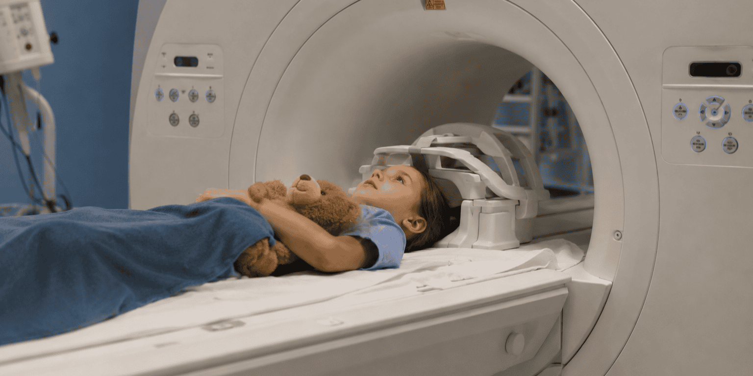
Pediatric MRI poses particular safety challenges to imaging facilities. Caregivers are often in the scanning room to comfort children, and sedation is frequently required to keep kids still during exams. 

  • But pediatric MRI safety hasn’t been studied as extensively as it has in adults, so researchers from five U.S. children’s hospitals reviewed MRI safety events that occurred at their facilities from 2017 to 2022.

Researchers focused on reported events that occurred within Zone IV, the area under the ACR’s four-zone safety model that includes the scanner room. They found…

  • A total of 146 safety events occurred in Zone IV out of 541k pediatric exams, for an event rate of 0.027%. 
  • An average of 4.9 events per year occurred at each site, or 3.3 events per 100k exams.
  • Event types involved projectiles (30%), burn/thermal injuries (13%), and implants (10%).
  • 78 events (53%) directly involved patients.
  • Ten events (6.8%) were classified as serious.

Frequent causes of events included medical equipment and supplies (anesthesia equipment and monitors, stethoscopes, and needles) and personal items like phones and badges.

  • Implanted devices like cochlear implants represent a growing challenge, as 20%-30% of children getting MRI scans have them, and safety events occurred despite sites following manufacturers’ guidelines. 

Why did the safety events happen? Study authors found that MRI safety protocols weren’t followed in 60% of events.

  • Lack of protocol adherence is a common refrain in MRI accidents that have involved adults, illustrating that all the guidelines and rules in the world won’t help if they aren’t followed to the letter.

The Takeaway

The new study on pediatric MRI safety highlights the fact that children shouldn’t just be treated like little adults when it comes to safe scanning procedures. The research offers a benchmark against which pediatric imaging facilities can measure themselves, while also offering additional guidance on mistakes to avoid when scanning kids.

Why MRI Providers Make Patient Comfort a Top Priority

Smart MRI providers know patient comfort is linked to profitability. After all, the comfort quotient impacts image quality, diagnostic accuracy, operational efficiency, and patient satisfaction. 

  • Yet at least one-third of adult patients report distress, moderate anxiety, and discomfort related to MRI, according to a systematic review involving 220 patients.

When patients are claustrophobic or uncomfortable, it negatively impacts MRI providers…

  • Fidgety patients lead to motion artifacts/poor quality images and the need to retake scans – impacting overall productivity.
  • Sedating patients requires additional safety precautions, costing time and money.
  • Cancellations and no-shows due to patient anxiety translate to lost revenue.
  • Patients who have an uncomfortable MRI experience may go elsewhere next time – reducing repeat business and word-of-mouth referrals.

But it doesn’t have to be this way. Providers should seek out high-performance MRI systems designed with patient comfort in mind…

  • Shorter scans – The shorter the scan, the easier it is for patients to stay still. Fujifilm’s ECHELON Synergy MRI features up to 50% scan time reduction over previous generation 1.5T MRI scanners, and a 70-cm-wide bore with a 62-cm-wide table to enhance comfort.
  • Software tools – A study found that 15-20% of MRI scans require re-scan due to patient motion. ECHELON Synergy offers software that mitigates the impact of motion artifacts, making it easier and quicker to complete scans on fidgety patients.
  • Open design – Put claustrophobic patients at ease and scanning needn’t stop mid-way through an exam. Providers are alleviating anxiety and boosting efficiency with Fujifilm’s OASIS Velocity MRI, which features a true open design, where patients have an unobstructed view for maximum comfort. OASIS Velocity also supports patients who weigh up to 660 pounds.
  • Easy access – Improving comfort for all patient populations can help keep a facility competitive. Fujifilm’s APERTO Lucent is a powerful permanent magnet open-sided 0.4T MRI that delivers an optimal patient experience. Its unique single-pillar design provides ideal technologist-patient access, and the wide, laterally moving table lowers to 20 inches, ensuring easy access for pediatric, elderly, and/or injured patients.

Hospitals and imaging facilities across the U.S. rely on Fujifilm for state-of-the-art, patient-centric MRI systems. For example…

Make patient comfort a top priority and give your MRI business a competitive edge with solutions from FUJIFILM Healthcare Americas Corp

Uneven Access to Brain MRI

Patients from disadvantaged neighborhoods or those traveling farther for brain MRI scans presented in worse clinical condition than patients with better access. That’s according to a new JACR study that reopens the debate over disparities in healthcare access. 

The past several years have seen numerous studies published that document disparities in healthcare access and their impact on clinical outcomes.

Many previous studies have also concentrated on access to care in rural areas, in which long distances make it harder for patients to travel to medical centers.

  • In the current study, researchers led by authors from Emory University flipped the script to examine care access in the Atlanta metropolitan area in an effort to quantify how distance and socioeconomic status might impact patient care. 

They examined the demographic backgrounds of 4.8k patients who got brain MRI scans over a one-year period starting in March 2019, calculating factors like distance from home to imaging facility and socioeconomic status based on the area deprivation index. 

  • They then correlated these data to patient illness severity – also known as acuity – when they presented for their scans, using a three-point scale ranging from normal (level 1) to findings requiring a change in patient management (level 3).

Based on the data, researchers found…

  • Patients in neighborhoods with lower socioeconomic status had 34% higher odds of level 2 acuity versus level 1 for inpatient scans and 27% higher for emergency scans. 
  • Patients living twice the distance from an imaging facility had 6.5% higher odds of level 2 acuity compared to level 1, and 15% higher for level 3.
  • Other factors affecting acuity level included age, race, and insurance status.
  • Medicaid recipients in particular were sicker, with 68% higher odds of acuity level 2 and 81% higher odds of acuity level 3 compared to those with commercial insurance. 

The findings track with other studies that have linked chronic health conditions with brain pathologies, such as the connection between diabetes and stroke. 

The Takeaway

The new findings offer additional details on how patient demographics affect both their health status and their access to care, in particular for advanced imaging scans like brain MRI. Follow-up studies could examine whether a similar phenomenon occurs with CT, which is the workhorse modality for emergency imaging. 

Why Radiology Leaders Are Turning to AI – And Why They’re Not Looking Back

From single-scanner clinics to university hospitals, radiology leaders around the globe face the same challenge: keeping up with rising patient demand while managing costs.

MRI volumes are climbing. Scanner hours and budgets? Not so much.

  • Under pressure to do more with less, decision-makers are reaching a conclusion that was unthinkable just a few years ago: AI-powered MRI is no longer a novelty – it’s a necessity.

No matter the size or scale of the operation, diagnostic imaging providers face a familiar set of challenges:

  • High capital costs – New scanners cost seven figures, and upgrades run hundreds of thousands.
  • Limited capacity – Most sites can’t easily add scanners, staff, or hours to meet demand.
  • Rising demand – MRI volume continues to grow as chronic conditions rise and preventive care gains traction.
  • Patient expectations – Long, uncomfortable exams frustrate patients who may look elsewhere.

AI offers a path forward, helping imaging teams handle more studies without compromising diagnostic standards.

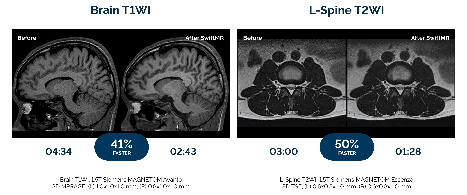
AIRS Medical built SwiftMR, AI-powered MRI reconstruction software, to meet today’s imaging challenges. Hospitals and clinics in over 35 countries use SwiftMR to:

  • Reduce scan times by up to 50% compared to standard protocols.
  • Deliver sharper images radiologists can trust.
  • Enhance the patient experience with shorter exams and fewer motion-related rescans.

SwiftMR is vendor-neutral, compatible with all MRI makes, models, and field strengths.

FDA-cleared, MDR-certified, and clinically validated, SwiftMR is trusted by over 300 imaging providers in the U.S. and over 1,000 globally, including:

These outcomes show that AI-powered MRI delivers tangible operational, clinical, and financial benefits across site types and geographies. 

Watch this video to learn more about SwiftMR.

The Takeaway

Radiology leaders are relying on SwiftMR to transform how they deliver care. From enterprise networks to single-scanner clinics, imaging teams are unlocking new levels of efficiency and patient care.

MRI of Bullet Fragments Is Possible

Radiology has a renewed focus on MRI safety following the tragic death of a New York man in an MRI accident last month. With that in mind, a new JACR study looks at adverse MRI events caused by an uncommon but still important phenomenon: retained bullet fragments in patients getting scans. 

MRI is radiology’s most powerful modality, but its strong magnetic fields can be hazardous – and on extremely rare occasions even fatal – for both patients and medical personnel.

  • Patients are supposed to be screened for metallic implants, jewelry, and other contraindications, but how often do providers know to ask about retained bullet fragments?

Having a retained bullet fragment on its own isn’t a contraindication for MRI, but providers do need to know where fragments are located and how large they are.

  • If pre-scan screening discovers a patient with a retained fragment, they typically receive X-rays of the involved area to determine location and size – scans should be aborted if the fragment is in a solid organ or within 5 mm of an important artery or vein.

If all these steps are taken and the scan goes ahead, how often do adverse MRI events occur? 

  • Researchers reviewed 6.1k X-ray reports that contained the terms “bullet” or “shrapnel” over 13 years, finding 284 patients who got an MRI scan after a retained fragment was found on radiography.

They found…

  • Only four patients (1.8%) experienced symptoms during MRI scans.
  • Each of the exams was terminated early due to patient discomfort, with three patients reporting burning and one general discomfort.
  • None of the symptomatic exams had the bullet in the MRI field of view.
  • No serious injury and no follow-up care was required. 

The Takeaway

The new findings are encouraging by showing that with careful patient screening and monitoring, MRI scans can be performed on patients with retained bullet fragments. But as always, MRI operators must remain vigilant and adhere to published MRI safety guidelines.

MRI Reveals Junk Food’s Toll on Carotid Arteries

As the U.S. government weighs a regulatory crackdown on ultra-processed food, a new study indicates the feds may be on to something. Researchers used MRI to discover that people who consumed more ultra-processed food had higher levels of carotid arterial plaque – a risk factor for cardiovascular disease. 

The FDA and the USDA on July 23 announced the start of a new initiative to investigate the risks of ultra-processed foods and their relationship to chronic diseases such as obesity, heart disease, and cancer. 

  • The project is widely seen as a priority of HHS Secretary Robert F. Kennedy, Jr. and his Make America Healthy Again movement. 

Meanwhile, arterial plaque buildup is a sign of atherosclerosis and has been linked to multiple clinical conditions, from stroke to intraplaque hemorrhage

In the new paper in American Journal of Preventive Cardiology, researchers noted the established association between adverse cardiovascular events and consumption of ultra-processed food and beverages, but the association with subclinical disease hasn’t been explored. 

  • So researchers reviewed carotid MRI scans of 768 participants from the Atherosclerosis Risk in Communities study, in which subjects also described their dietary intake with a 148-item questionnaire. 

MRI scans were correlated with dietary habits, finding that compared to the lowest quartile, people in the highest quartile of ultra-processed food consumption had…

  • Greater total arterial wall volume. 
  • Greater total lipid core volume and maximum lipid core area.
  • Higher maximum segmental wall thickness.

No correlation was found between arterial plaque and other types of diet measures, such as carbohydrate and fat intake or glycemic load index.

  • The findings suggest that much of the negative health effect from ultra-processed foods comes from their contribution to arterial plaque buildup, which could occur through their unfavorable nutrient profile leading to alterations in blood lipids.

The Takeaway

In today’s hyperpolarized political environment – in which scientific inquiry is often subordinated to already-solidified beliefs – the new findings connecting MRI measurements of carotid artery plaque to ultra-processed foods offer a foundation for public policy changes that could indeed improve the health of Americans.

MRI Accident Turns Deadly

A tragic MRI accident in Long Island, New York, has turned deadly. A man who was pulled into a mobile MRI scanner by a heavy chain he was wearing died of his injuries. 

Keith McAllister was waiting outside a mobile MRI trailer operated by Nassau Open MRI on Long Island as his wife received a knee scan.

  • McAllister was wearing a weight-training chain around his neck that weighed some 20 pounds.

When he entered the trailer to help his wife get off the scanner table, the system’s powerful 1.5T magnetic field drew him against the magnet. It took staff an hour to free him.

Investigators are still looking into the details of the episode, but it underscores the shortcomings in how MRI safety is regulated in the U.S., where fatal MRI accidents are extremely rare but still do occur.  

  • That’s according to MRI safety expert Tobias Gilk, vice president at architectural firm Radiology Planning and founder of Gilk Radiology Consultants, who spoke to The Imaging Wire about the accident.

The U.S. has some of the most comprehensive and sophisticated guidelines on MRI safety, encapsulated in the ACR Manual on MR Safety.

  • What’s more, the radiology community including ACR, ISMRM, ASRT, and others are currently observing their annual MR Safety Week to promote safe MRI scanning – an event that started just a few days after McAllister died.

But despite the great leaps in knowledge about MRI safety, Gilk believes that keeping patients safe is complicated by the exponential growth in the modality’s complexity, while actual enforcement of safety standards is lacking. 

  • Many state health departments don’t even address MRI safety as they focus more aggressively on regulating ionizing imaging modalities like CT and X-ray, and healthcare certification bodies like the Joint Commission lack enforcement teeth.

Instead, MRI safety often becomes the responsibility of technologists who frequently must juggle multiple tasks as they manage both patients and scanner operations.

  • This can be particularly challenging in mobile MRI coaches, often staffed by a single MRI technologist where the only barrier between the outside world and the scanning environment is just a single – often unlocked – door. 

The Takeaway

The tragic death of Keith McAllister in a mobile MRI trailer shows that all the guidelines and safety events in the world won’t keep patients safe unless accompanied by stronger enforcement of the knowledge the radiology community already has. We can do better.

Get every issue of The Imaging Wire, delivered right to your inbox.