GE HealthCare this week announced FDA clearance for Photonova Spectra, the company’s first photon-counting CT scanner. While GE isn’t the first vendor with a commercially available PCCT scanner, it’s hoping to differentiate the system by highlighting the combination of ultrahigh-resolution scanning with spectral imaging.
Photon-counting CT represents a huge leap forward in CT instrumentation that’s not only driving new clinical applications but is also helping radiologists perform routine CT exams with better resolution and lower radiation dose.
- PCCT scanners directly convert photons to digital data, instead of using conventional CT’s two-step energy-integrating technique, resulting in images with less noise and supporting acquisition protocols with lower radiation dose.
Siemens Healthineers brought the first photon-counting CT scanner to market with the 2021 FDA clearance of Naeotom Alpha.
- Since then, Siemens has had the market for whole-body PCCT to itself, with only niche photon-counting scanners getting FDA clearance.
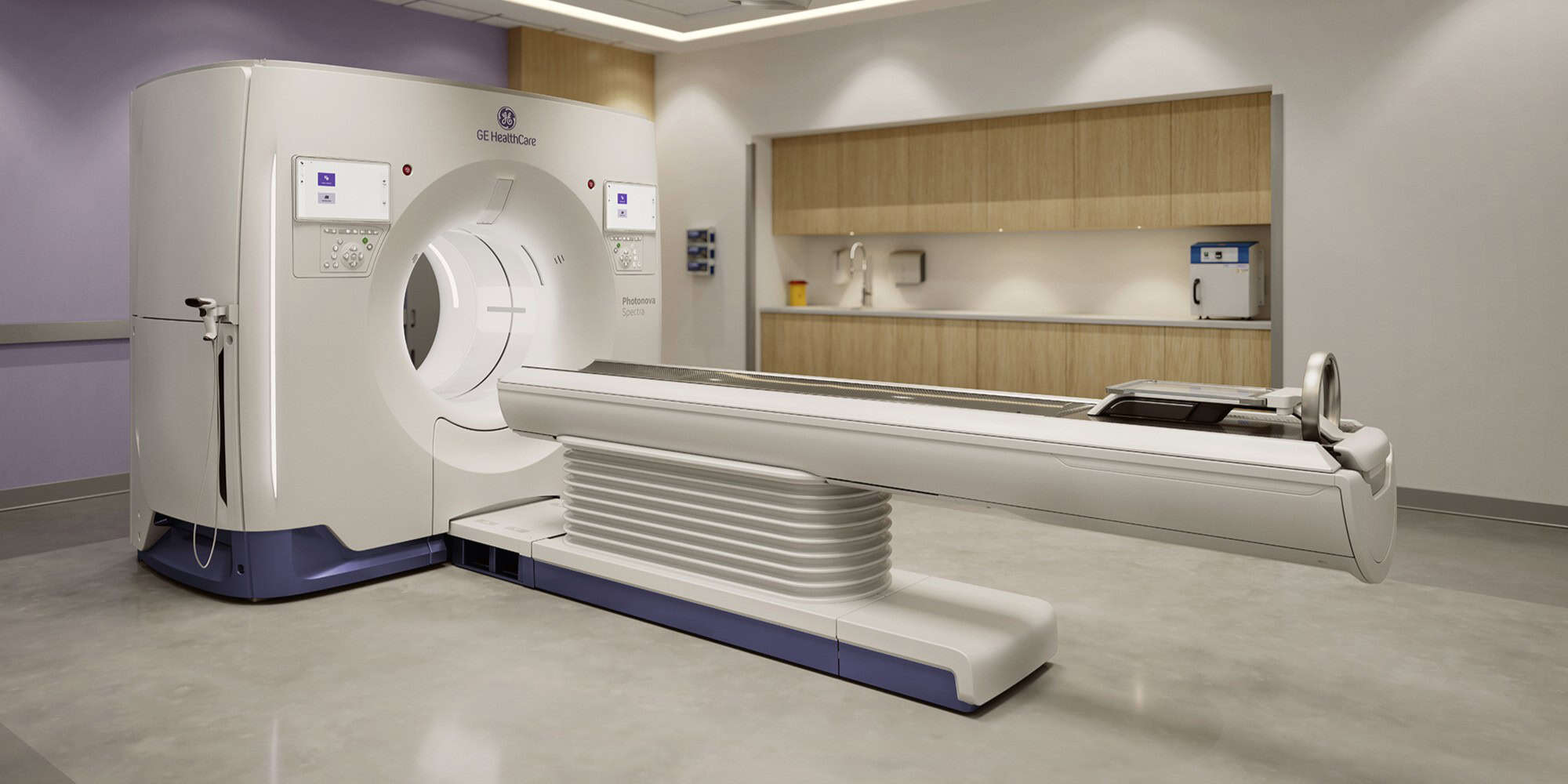
But we’re here to talk about GE’s Photonova Spectra, so let’s get to it. The system is based on GE’s Deep Silicon detector technology, which uses a novel semiconductor detector material that’s particularly suited for spectral imaging.
- Spectral CT acquires images at different energy levels, which is useful for detecting disease because malignant and benign tissue respond differently to different energy spectra.
GE is highlighting Photonova Spectra’s 8-bin energy resolution, which means the scanner separates incoming photons into eight distinct energy ranges – or bins – rather than grouping them into one or two.
- This enables Photonova Spectra to deliver much more precise spectral imaging than previously possible, with better quantitative accuracy and improved differentiation between materials like bone and soft tissue, according to GE CT executive Chad Rowland.
Spectral CT has developed a reputation as a technology that’s powerful but complex, and GE addressed this issue with workflow tools that make spectral imaging “always on” and easier than ever to perform.
- GE is banking on the combination of spectral imaging with Photonova Spectra’s ultrahigh-resolution images being a game-changer for many sites considering adopting their first PCCT scanner.
The Takeaway
FDA clearance for GE HealthCare’s Photonova Spectra photon-counting CT scanner is great news for the vendor that puts it on a level competitive footing with Siemens as a CT innovator. But it’s also good news for imaging providers, giving them another option for delivering to patients the benefits of PCCT – lower radiation dose and better image quality.


