Measuring AI’s Impact in Real-World Radiology

One of the criticisms of AI in radiology is that there isn’t much data on its effectiveness in real-world environments. Well, a new study in JACR aims to fill that gap by detailing how AI adoption at a large imaging network in Europe affected key radiology performance metrics. 

Plenty of studies have been published on AI’s positive contribution to radiologists’ diagnostic performance. 

  • But in most cases this research was performed under controlled conditions, or involved a single AI algorithm working on a particular task. That’s left many wondering how well AI would perform under actual conditions. 

In the new study, researchers tracked the performance of 10 AI algorithms from seven vendors that were implemented over the Incepto Medical AI orchestration platform at 3R Swiss Imaging Network, a 20-center outpatient radiology network in Switzerland. 

  • Over 4.5 years, researchers tracked the network’s performance before and after AI implementation for the major radiology key performance indicator, report turnaround time (TAT). They also tracked AI adoption rates and infrastructure latency, or how delays in data transfer between PACS networks affected AI usage.

In all, 389k AI-assisted imaging exams were processed over the implementation period from 2021 to 2025, with researchers finding…

  • AI’s biggest impact was in high-volume modalities, reducing TAT for trauma radiography (-26%) and knee MRI (-18%).
  • AI improved the network’s diagnostic capacity, performing the work of almost half a full-time-equivalent radiologist for trauma radiography alone (0.46 FTE). 
  • AI usage rates were high, with 91% reporting active AI adoption and 66% reporting regular use.

On the downside, latency in data transfer between PACS networks slowed down AI performance, and in some cases AI results arrived after reports were finalized – too late to make a clinical impact…

  • Median total latency was 2.06 minutes per exam, with 72% attributable to data routing and tasks like fetching studies from PACS.
  • AI’s “too late rate” was 7.2% overall, ranging from 13% for chest CT to 3% for knee MRI, with 6.8% for trauma X-ray.

How did the network’s radiologists feel about AI? Researchers surveyed over 50 radiologists working for the network, finding that most of the differences in before-and-after opinions weren’t statistically significant. 

  • The only difference was a modest gain in the perception that AI made radiologists more productive (from 2.57 to 2.94 on a five-point Likert scale).

The Takeaway

The new study fills in a major gap in real-world experience regarding radiology AI implementation. It also reveals areas for improvement, as slow data transfer rates between networks can prevent AI results from reaching radiologists in time. 

One-Stop CT Chest Screening

Could opportunistic screening be driving the evolution of low-dose CT from a screening test just for lung cancer into a broader one-stop exam for multiple chest pathologies? A new study in JACR suggests as much – and provides a blueprint for how to do it. 

There’s no question LDCT lung cancer screening is gaining traction around the world. But as screening programs are starting, clinicians are finding that CT scans can detect far more than just lung cancer. 

  • Emphysema, commonly associated with COPD, is one such condition, as it goes hand-in-hand with lung cancer in people with long smoking histories. Another is coronary artery calcium (CAC), a known risk factor for cardiovascular disease. 

In the new study, researchers wanted to investigate the prevalence of COPD and CAC in their LDCT screening program covering three hospitals in the NewYork-Presbyterian system. 

  • In addition to documenting prevalence rates, they wanted to connect clinical outcomes to social determinants of health, which have been demonstrated to affect patient care. 

They tracked results from 2.4k people who got baseline LDCT scans from 2015 to 2022, finding…

  • Emphysema was detected in 77% of patients, and CAC in 65%.
  • 87% of patients with emphysema had no previous diagnosis.
  • The proportion of patients getting pulmonary care after their LDCT scan increased (from 24% to 46%), as did the rate of those getting cardiovascular care (from 43% to 63%).  
  • Lower household income was associated with 57% higher adjusted odds of emphysema, and food insecurity with 52% higher odds. 
  • No social determinants of health were associated with CAC burden.

It’s worth noting that the study did not use any IT tools to classify COPD and CAC, like AI image analysis or longitudinal tracking and reporting. 

  • It’s entirely possible that the results could be even more promising given the plethora of algorithms that have become available for opportunistic screening applications. 

The Takeaway

The new study on incidental CAC and emphysema findings on LDCT exams shows that lung screening has the potential for opportunistic disease detection even if you don’t have fancy AI algorithms at your disposal. All you need are radiologists and clinical staff dedicated to providing better patient care. 

Repeat MRI Scans Take Operational Toll

Nobody likes having to repeat a task, and nowhere is that more evident than in MRI. A new study in JACR quantifies the operational toll of MRI scans that have to be repeated due to patient motion, revealing both good news and bad news. 

Despite its unquestioned diagnostic power, MRI is the imaging modality most susceptible to motion artifacts.

  • That’s because patients have to lie motionless for long periods of time in a confined, noisy metal tube – an environment tailor-made to induce claustrophobia. 

MRI vendors have introduced a variety of new technologies and pulse sequences to speed up exams, but patient motion is still a nagging problem. 

  • To investigate the issue further, researchers from NYU Grossman School of Medicine collaborated with Siemens Healthineers to document MRI exam repeat rates over six months at NYU.

In all, 85.3k MRI exams were included in the analysis, with the following findings…

  • 4.8% of all MRI exams (4k) included at least one repeated sequence.
  • The highest repeat rate occurred in pediatrics and the lowest in breast MRI (9.9% and 3.5%, respectively). 
  • Repeat acquisitions added up to 115 hours of scanner time, equivalent to 268 lost MRI scanning slots. 

To take the study deeper, researchers more closely analyzed brain MRI and MR cholangiopancreatography (MRCP) studies, finding…

  • For 77 patients undergoing brain MRI, repeat acquisitions had better image quality and diagnostic confidence, but 20% of repeat sequences did not improve. 
  • For 27 patients undergoing MRCP, repeat acquisitions were less successful, as they didn’t improve image quality or diagnostic confidence.

So that’s the bad news. What’s the good news? The study’s 4.8% repeat rate was actually lower than those reported in previous research into motion-related repeats, which have ranged from 7.5% to 30%. 

  • One reason for the discrepancy is that the current study relied on exams labeled as repeats by technologists, which could have resulted in an underestimation. 

But another reason is the fact that MRI practices have adopted a wide range of techniques and technologies that reduce the impact of patient motion, like shorter protocols, parallel imaging, compressed sensing, and deep learning reconstruction. 

The Takeaway

The new findings into the operational toll of MRI motion artifacts provide useful insights into the extent of the problem. But they also include a silver lining – that the modality’s relentless pace of technological innovation could make repeat exams less of an issue in the future. 

What Now for the USPSTF?

After not meeting for over a year and undergoing major turnover, HHS Secretary Robert F. Kennedy, Jr. last week announced new members of the U.S. Preventive Services Task Force – including the USPSTF’s first-ever radiologist. 

USPSTF was created in 1984 as a panel of experts tasked with producing evidence-based advice on preventive medical care, like cancer screening guidelines. 

  • But it’s that last responsibility that’s drawn the USPSTF into controversy. The group in 2009 changed its blanket recommendation regarding screening mammography for women aged 40 to 50, instead recommending women consult with their doctors. This sparked a backlash that persisted until 2023, when it said it would lower the age back down to 40. 

Indeed, the USPSTF has often been accused of being too conservative in recommending new technologies that have life-saving potential. 

  • For example, its implementation of the new mammography screening guidelines neglected to recommend supplemental dense breast screening, despite the demonstrated connection between dense breasts and cancer risk.

Enter RFK, Jr., who as HHS secretary has appointment authority over the panel’s 16 members (who, it should be noted, serve as volunteers rather than as federal employees).

  • He’s held the USPSTF with the same suspicion he reserves for the rest of the U.S. medical establishment, and on his watch the group has gone a year and a half without a meeting. He also removed both of the panel’s vice chairs in May. 

RFK, Jr. hadn’t revealed much about his intentions for the panel, but that ended on September 17, when HHS announced a new slate of appointments. 

  • He tapped eight new members, including a number of medical specialists, addressing a frequent criticism that the USPSTF was too heavily weighted toward primary care and lacked specialty experience. 

And in what is a first, he named a radiologist to the USPSTF: Dennis Wulfeck, MD, of MBB Radiology, a Radiology Partners-affiliated practice in Jacksonville, Florida. 

  • Wulfeck earned his MD at Wright State University, performed his residency at the University of Louisville, and completed a fellowship in nuclear medicine at the University of Cincinnati. Wulfeck has published research in nuclear medicine and has been involved in radiology practice issues. 

What now for the USPSTF? While the new appointments seem to represent physicians from U.S. medicine’s mainstream, their work will be closely scrutinized for signs of politicization, particularly around RFK, Jr.’s pet issue of childhood vaccines. The USPSTF hasn’t said when its next meeting will take place. 

The Takeaway

Radiology has long had a complicated relationship with the USPSTF, mostly due to its conservative stances on cancer screening and the adoption of new imaging technologies. Will that change with a radiologist now serving on the panel? We shall see. 

Expanding CT Lung Screening at WCLC 2026

Low-dose CT lung cancer screening appears set to be the next major population-based cancer screening exam. But much work remains to make LDCT screening available to as many eligible people as possible while optimizing scanning protocols.

That’s according to sessions at IASLC’s World Conference on Lung Cancer (WCLC 2026), which just wrapped up in Seoul, South Korea. 

  • WCLC 2026 sessions ranged from expanding scanning to people at high risk but without smoking histories to using AI to not only detect and characterize lung nodules but to predict future cancer risk.

Some of the key findings from WCLC 2026 included…

  • Australia started its national LDCT screening program in 2025, and researchers expect a national-level stage shift to earlier diagnosis. At a 50% participation rate, stage I diagnoses should grow from 32% currently to 42% for women and 43% for men. 
  • The Ready to Screen (R2S) trial in Australia found that of 1.7k eligible screening candidates surveyed, 85% “definitely” intended to get screened and 26% said they had already received an LDCT scan. 
  • Taiwan’s national screening program showed that – at least in East Asia – people with a family history of lung cancer should be screened. Of 2.8k cancers detected, there was a 74% higher detection rate in those with family histories versus smoking histories (18 vs. 10 cancers per 1k screened), with more early-stage cancers detected (92% vs. 83%).
  • An LDCT screening program in China screened 6.7k people – including those with risk factors besides smoking – finding a 56% lung cancer mortality reduction.  
  • French clinicians successfully added smoking cessation therapy to their LDCT program, with 88% screening attendance and participants 44% less likely to smoke.
  • MIT’s Sybil AI algorithm was more accurate than Lung-RADS in predicting one-year cancer risk from suspicious nodules, with higher AUC when applied to two large lung screening trials (NLST and P-IELCAP). 
  • Researchers found that applying Sybil to coronary artery calcium scans could also predict lung cancer risk for both smokers and non-smokers over a 15-year follow-up period.
  • Researchers used Softek Illuminate’s Illuminate AI software for identifying incidental pulmonary nodules as a complement to an LDCT screening program, finding that the algorithm discovered more lung cancer cases than conventional screening. 

The Takeaway

This week’s WCLC 2026 sessions point out the rapid progress being made around the world in expanding access to LDCT lung cancer screening – as well as the role that AI-based software tools can play in making screening more effective and more widely accessible. 

Top 10 AI Vendors by FDA Approvals

The FDA updated its list of regulatory authorizations for AI-enabled medical devices, covering approvals granted through the end of June 2026. The new numbers show radiology maintaining its strong lead over other medical specialties. 

The FDA issues quarterly updates on marketing authorizations for AI-enabled products, a category that includes dedicated software applications as well as AI-enhanced hardware, like scanners with onboard AI functionality. 

  • The list is closely watched by healthcare industry observers, and radiology has typically secured more than three-quarters of the authorizations – not surprising given the specialty’s image-intensive nature. 

The most recent list tracks FDA actions through the end of June 2026, indicating that the FDA has…

  • Authorized 1,614 AI-enabled medical devices since it started keeping track, up 5.9% from Q1 2026. 
  • Authorized 1,230 radiology devices, or 76% of all AI-enabled medical authorizations. 
  • In the second quarter of 2026, the FDA authorized 89 AI-enabled medical devices, slightly fewer than the 92 devices it authorized in the first quarter of 2026. 
  • For the quarter, 66 authorizations (74%) were for radiology devices, similar to the Q1 ratio (75%).
  • GE HealthCare maintained its AI authorizations lead with 134, thanks in part to approvals secured by companies it has acquired over the years. 
  • Next are Siemens Healthineers at 101, then Philips at 62, Canon at 51, United Imaging at 45, Aidoc at 34, and DeepHealth at 32, with all numbers including acquisitions. 
  • Rounding out the top 10 are Samsung (21), RapidAI (20), and Hyperfine (13).

The Takeaway

The new numbers track pretty closely with data on AI-enabled regulatory authorizations the FDA released for the first quarter. It will be interesting to see whether the agency’s pace changes as it comes to a final decision on regulating generative AI algorithms used for direct clinical care – a task for which it is currently soliciting feedback. 

Harrison’s Teleradiology Play

AI developer Harrison.ai is expanding its business model by supporting a teleradiology venture called Frontier Radiology that’s scaling up and hiring radiologists. But the move drew unwanted attention from a critical article in the Australian news media this week.

Harrison is best known for its suite of AI algorithms for radiology applications, as well as its Harrison.Rad 1.5 foundation model that can draft reports for radiologist review.

  • Harrison is based in Australia, but has recently made the U.S. market a major focus, with co-founder and managing director Dimitry Tran relocating to California to spearhead the initiative.

But it was Harrison’s newfound U.S. focus that apparently drew the attention of ABC News, an Australian news outlet that on September 7 published a critical article that questioned the company’s moves, in particular its involvement with Frontier Radiology.

  • One bone of contention in the story was whether Harrison’s U.S. emphasis was appropriate given the Australian government’s 2025 investment of US$23M in the company. The story also claimed Harrison was planning layoffs of Australian staff and that its integration of AI with Frontier’s clinical services could be a conflict of interest.

But in an interview with The Imaging Wire, Dimitry Tran clarified Harrison’s strategic direction and its involvement with Frontier Radiology.  

  • Tran noted that Frontier is owned and operated by a radiologist – Joshua Ewell, DO – in accordance with U.S. rules requiring physician ownership of entities providing clinical services. 

Ewell’s LinkedIn profile says Frontier will be an “AI-native radiology group” built entirely around AI foundation models.

  • Harrison is providing Frontier with non-clinical services, including its suite of AI algorithms, which are already helping Frontier teleradiologists achieve industry-leading report turnaround times. But Frontier radiologists are free to use any AI solutions they wish – including those of Harrison’s competitors. 

Tran noted that close cooperation between AI developers and imaging services providers is hardly unusual anymore in radiology. 

  • Indeed, two of the specialty’s largest U.S. providers – Radiology Partners and RadNet – have formed their own AI divisions to provide algorithms to both their own radiologists and outside customers. 

The ABC News article conflated a number of recent developments into a narrative that doesn’t reflect reality, Tran believes. 

  • For example, the layoffs that occurred earlier this year at Harrison were connected to the company’s transition from an aggressive R&D phase into a commercialization push and weren’t related to Harrison’s U.S. entry.

And the Australian government’s funding was part of an investment that gave it a single-digit equity stake in the company – a stake it retains to this day and that will prove increasingly profitable with Harrison’s growing success.

  • Even as it supports Frontier, Harrison plans to continue its focus on AI algorithm development and commercialization, especially of the Harrison.Rad foundation model – while keeping the “vast majority” of its employees in Australia. 

The Takeaway

Putting aside the ABC article’s negative spin, Harrison’s move into teleradiology offers an intriguing twist on the growing integration between AI and imaging services providers. Given ongoing workforce shortages and rising imaging volume, it’s perhaps the best way to move the chains toward finding relief for beleaguered radiologists. 

VA Radiologist Exodus ‘Tip of Iceberg’

A “mass exodus” of radiologists from the VA’s medical center in Washington, DC, represents just the tip of the iceberg for the health system’s radiology personnel woes. Other VA centers are reported to have seen similar departures, and the VA’s teleradiology program appears to be buckling under the strain. 

News about radiologist staffing problems in the VA came to light in July with the publication of news articles claiming that the Trump administration’s 2025 ban on remote work for federal employees had prompted most radiologists at the VA Washington DC Medical Center to quit.

  • While the ban was later rescinded for radiologists, the edict reportedly “spooked” many radiologists who were already chafing under VA salaries that were far lower than what they could find in the private sector.  

An investigation by the VA’s Office of Inspector General confirmed the Washington, DC, medical center’s problems, documenting the departure of four of the facility’s five full-time radiologists as well as its chief of radiology.

  • But the question remained – how widespread are the VA’s radiologist staffing woes, and have the same problems occurred at other VA medical centers?

Indications are that it’s a growing problem. A VA employee who contacted The Imaging Wire reported that VA radiology departments around the U.S. “got decimated,” with some losing 50% of their radiologist staff.

  • This includes VA medical centers in Houston, Dallas, and San Antonio, and a number in Florida. 

Interestingly, the source claims that the return-to-office edict has only been a “minor” contributor to the exodus, instead attributing the departures to a vicious cycle of lower salaries and higher workload. 

  • The VA’s salary cap of $400k simply isn’t competitive with private-sector radiology, where a recent compensation survey pegged average U.S. radiologist compensation at $610k – a 53% difference. 

The shortage of in-person radiologists is prompting the VA to lean more heavily on its National Teleradiology Program, an operation headquartered in Menlo Park, California, that employs dozens of radiologists to interpret imaging studies overnight and on weekends.

  • But NTP is facing a personnel exodus of its own, especially after NTP officials required daytime radiologists to pick up the slack by also working nights and every other weekend. The source estimated that, at its peak, NTP had about 120 radiologists. That number now stands at 62.

What impact are the departures having on VA patient care? The VA mandates a one-hour turnaround time (TAT) for STAT imaging exams. 

  • But the source claims that TAT has ballooned to as high as 10 hours. “Imagine not getting critical diagnoses for ER patients until 10-12 hours later!” the source said.

The Takeaway

The new revelations suggest that the problems reported at the VA Washington DC Medical Center are actually endemic throughout the VA system. And given the growing gap between VA and private radiologist salaries, they aren’t likely to recede anytime soon, regardless of the agency’s work-from-home policies.

PET Tracer for Cardiac Amyloidosis Shows Promise

New results were presented this weekend on a PET radiopharmaceutical being developed by Bayer for imaging cardiac amyloidosis. The tracer turned in promising results in a paper presented at ESC 2026 and published simultaneously in JAMA.

Cardiac amyloidosis occurs when abnormal proteins called amyloid fibrils build up in heart tissue, eventually leading to heart failure and death. It was once thought to be untreatable, but new pharmaceutical therapies have become available that slow disease progression and improve patient outcomes. 

  • But first you have to make a diagnosis, and that’s been the hard part. Multiple imaging modalities are often required, and patients with cardiac amyloidosis often experience diagnostic delays of one to two years or more.

Enter Bayer, which earlier this year acquired a pair of radiopharmaceuticals from Attralus, one for PET imaging and the other for SPECT. The PET agent is iodine-124 evuzamitide, a radiolabeled synthetic peptide that binds to the amyloid fibrils that characterize cardiac amyloidosis. 

  • The acquisition was a bit of a departure for Bayer, which until the purchase had focused its imaging business mostly on contrast agents for X-ray, CT, and MRI. 

But the results presented over the weekend from the Phase III REVEAL trial indicate Bayer may have placed a good bet. 

  • Researchers enrolled 170 patients from 18 U.S. centers who got PET/CT scans with iodine-124 evuzamitide. Three physicians with experience in cardiac PET/CT reviewed the results.

The REVEAL investigators discovered…

  • Iodine-124 evuzamitide had 94% sensitivity for cardiac amyloidosis, with 86% specificity. 
  • Positive predictive value was 85%, and negative predictive value was 94%.  
  • The radiotracer showed high diagnostic accuracy for both types of cardiac amyloidosis, light chain (AL) and transthyretin (ATTR) amyloidosis.
  • The radiotracer was well tolerated by patients. 

The findings indicate that iodine-124 evuzamitide has potential “as a reliable first-line test to exclude cardiac amyloidosis,” meaning it could rule out the need for invasive biopsy. 

  • What next for the tracer? Bayer said it would be discussing the REVEAL data as well as regulatory submissions with the FDA and other health authorities.

The Takeaway

New results from REVEAL indicate that Bayer’s iodine-124 evuzamitide PET radiotracer has the potential to fundamentally change the diagnosis of cardiac amyloidosis, historically one of the more challenging heart conditions to detect and manage. 

Radiology Salary Growth Beats Other Specialties

Radiologists enjoyed faster salary growth than physicians overall in the latest salary survey from Doximity. Radiologists also moved up several positions on the list of highest-paid doctors. 

Doximity operates a widely used digital platform that counts around 85% of U.S. physicians as members. 

  • The company’s annual salary survey is a closely watched barometer of physician compensation, as well as other trends like compensation by metro area and physician specialty. 

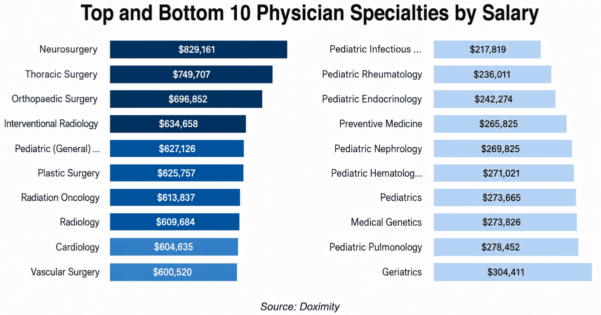
The 2026 survey found that radiologists had an average annual salary of $610k, up 6.6% compared to $572k in the last survey. Radiology’s growth exceeded the 2% average growth rate for all physician specialties. 

  • Radiology moved up three positions to occupy the #8 slot among highest-paid specialties, compared to #11 in last year’s survey. 

Despite radiology’s progress, salary growth in the specialty actually slowed a bit compared to Doximity’s 2025 survey, when it rose 7.5%. 

  • Back then, radiology had the fourth-fastest salary growth among medical specialists. 

Count yourself lucky if you’re an interventional radiologist. The field saw the largest increase in annual compensation among all physician specialties – 10.8%, bringing average compensation to $635k.

Besides just compensation, the 2026 survey also queried physicians about other employment-related topics, finding…

  • 85% reported that the physician shortage had affected their clinical practice.
  • 76% said the shortage and other pressures had compromised the quality of care they deliver.
  • The gender salary gap remained at 26% for the second straight year as men made an average of $122k more than women.
  • At 10%, radiology actually had one of the smallest gender pay gaps.

The survey also drilled down into physician attitudes toward artificial intelligence, which is being proffered as a potential solution to the workforce shortage, finding that…

  • 66% reported using AI on a daily or weekly basis.
  • 23% expected AI to boost their compensation within the next year.
  • 67% thought that doctors who stay current with AI tools will have an earnings advantage over physicians who don’t.

The Takeaway

The new Doximity physician survey showed that despite workload challenges, radiology remains one of the best-compensated physician specialties. It also highlights physicians’ changing attitudes toward AI, as what was once seen as a competitive force is now viewed as a complementary and even beneficial technology for doctors.

Get every issue of The Imaging Wire, delivered right to your inbox.