AI Reduces Mammography Workload

Using AI to triage low-risk breast screening exams that don’t need extra review could remove more than three-quarters of mammography cases from radiologists’ workload and allow them to spend more time on high-risk cases. That’s according to a new study in Radiology: Artificial Intelligence that confirms other recent studies. 

Much of recent mammography AI research has focused on its ability to triage low-risk cases to avoid additional radiologist review – saving precious personnel resources.

  • This is particularly valuable in Europe, which uses a double-reading paradigm in which two radiologists review all mammography cases (the U.S. employs single readers but tends to screen women annually rather than every two years). 

The new study comes from France, which employs a slightly different paradigm from the rest of Europe. Double reading is conducted only for lower-risk BI-RADS 1 and 2 cases, while BI-RADS 3-5 go directly to diagnostic workup. 

  • As such, double reading occurs with cases that have low cancer prevalence, which can make it more difficult for radiologists to detect cancers that don’t occur very often.

But what if you offloaded low-risk double reading to AI? 

  • In the new paper, researchers tried that with Therapixel’s MammoScreen AI algorithm, which was employed retrospectively to analyze mammograms from 42.4k women acquired from 2015 to 2019.

AI results were compared to standard radiologist double reading, with the following findings…

  • AI classified 77% of cases as low-risk, meaning these could be safely triaged from the double-reading paradigm.
  • AI missed only one cancer in the low-risk group, a rate the researchers characterized as “small but measurable.” 
  • Eleven cancers were found in the group AI classified as non-low-risk, which would have undergone double reading anyway in the AI triage paradigm.
  • Rates of interval cancer (cancer that occurs between screening rounds) were 5X higher in the cases AI classified as non-low-risk compared to low-risk (2.16 vs. 0.47 cancers per 1k exams). 

Using AI to classify and remove low-risk cases from double reading could therefore save significant resources from the French mammography screening program, with a “small but non-zero risk” of missed cancers.

The Takeaway
The new results track with findings from other recent studies that apply AI to mammography screening, particularly in Europe. While the French reading paradigm is unique, it’s instructive to see that AI maintains its ability to reduce radiologist workload across different types of breast cancer screening programs.

Does AI Still Scare Off Radiology Trainees?

Is AI still scaring off medical students from picking radiology as a specialty? A new study in Academic Radiology found that while prospective radiology trainees don’t seem as worried as they were after radiology AI burst onto the scene in 2015, they still have concerns about how AI will affect the profession. 

Radiology has long been seen as the medical specialty most at risk of broader AI adoption, largely because early AI applications focused primarily on image analysis.

  • These fears led to a widely publicized dip in radiology residency applications after 2016, the year after IBM Watson debuted at the RSNA show and when AI guru Geoffrey Hinton, PhD, issued his famous advice to stop training radiologists. 

But interest in radiology rebounded shortly after that. AI adoption was slower than anticipated, and few hospitals have proven willing to turn over radiologists’ duties to computers. 

  • Given the changes, how have the attitudes of medical students toward AI evolved in the last 10 years? Researchers decided to survey Canadian medical students and residents to find out.

In all, 401 respondents replied to the survey, of whom 13% had ranked radiology as their top specialty choice, with the following findings…

  • Only 2.5% said AI was “extremely influential” in affecting their specialty choice, with 57% saying it had a “slight/moderate impact” and 35% stated “no impact.”
  • AI was more important for those ranking radiology in their top three, with 91% saying AI influenced their decision compared to 54% of those uninterested in radiology. 
  • For those interested in radiology, 33% said AI made them feel discouraged, 13% were encouraged, and 33% reported no AI influence.
  • Those who believed AI would reduce radiologist demand were 50% less likely to be interested in a radiology career.

How to interpret the results? The authors felt the findings showed that AI had either no influence or a slight/moderate effect on specialty choice, but the impact was greater in those who were interested in radiology. 

  • They also saw a “growing polarization” among trainees, in that while many viewed AI as a threat to their job security, some saw it as an opportunity for innovation. 

The Takeaway

Medical students have complex and nuanced attitudes toward AI in radiology, as the new study indicates. But the findings suggest that past fears of radiology AI have evolved into a more measured view that better reflects real-world AI adoption.

AI for PE Detection: ‘Selective but Meaningful’

AI made a “selective but meaningful” contribution to radiologist interpretations of CT pulmonary angiography scans for pulmonary embolism. The study, published in Radiology: Artificial Intelligence, offers valuable insights into real-world implementation of AI on a large scale. 

One of the major criticisms of AI is that algorithms used in real-world clinical situations don’t perform as well as they do in the controlled environments that vendors use to acquire data for regulatory submissions.

  • AI performance can drop off as much as 20 to 30 percentage points for important metrics like sensitivity and specificity. 

The new study sought to investigate this phenomenon by analyzing a real-world implementation of Aidoc’s AI algorithm for PE detection. 

  • Researchers assessed the algorithm’s performance for analyzing CTPA exams across a variety of clinical environments in an integrated health network, including the emergency department and inpatient and outpatient settings. 

Scans of 29.5k patients acquired from 2021 to 2023 were included. AI analyzed images in real time, after which exams were interpreted by radiologists who knew the AI findings. Researchers found…

  • Radiologists using AI had higher sensitivity than the algorithm on its own (99% vs. 85%).
  • Specificity was more or less the same (99.8% vs. 99.5%).
  • Agreement between radiologists and AI was high (98%).
  • Agreement was higher when AI assessed cases as negative rather than positive (98% vs. 94%).
  • Radiologists disagreed with AI in 2.2% of cases. The final determination by a panel of expert thoracic radiologists strongly favored radiologists (89%).
  • Of the 3.3k cases positive for PE, 0.81% were detected only by AI – or 26 cases.

In analyzing the results, the researchers characterized AI’s contribution as “selective but meaningful.”

  • AI-positive results meant scans might require more scrutiny from radiologists, while an AI-negative call might be supportive – but not definitive – for negative PE.

The Takeaway

The new study of AI for PE detection is a fascinating look at real-world AI deployment. While the sensitivity, specificity, and agreement numbers are interesting, what draws our attention is the 26 PE cases caught only by AI over 18 months of use. That boils down to 26 patients whose clinical condition wasn’t missed, and 26 potential malpractice lawsuits that were never filed.

AI’s ROI Paradox

As radiology AI slowly moves from pilot projects to widespread clinical adoption, a new survey reveals a paradox: The technology is popular with radiologists, but few imaging facilities using AI have collected hard data showing its return on investment.

AI’s slow clinical adoption has frustrated both clinicians and algorithm developers alike, but the technology is gaining steam.

  • Despite growing clinical evidence, research on AI’s financial value and ROI has been slower in coming. 

To remedy that situation, AI governance startup Croviz.ai conducted a study of 445 radiology AI users on the economics and evaluation of AI. The full report is available here.

  • Survey respondents came from 12 different countries and included a variety of professional roles, including vendor executives, radiologists, and IT and informatics personnel.

Croviz founders Ayman Talkani and AadilMehdi Sanchawala found that while radiology AI power users loved the technology – and some refused to work without it – few had determined a positive financial return from it. Findings included…

  • 95% of sites already using AI had renewed at least one contract with an AI vendor in the last 12 months.
  • But only 30% had quantified a positive financial ROI from AI.
  • 54% cited better quality of life for radiologists as their main reason for renewing an AI contract.

So if AI’s value hasn’t been demonstrated, why are radiology sites renewing AI contracts?

  • The number one reason cited by 54% of those renewing contracts was because their radiologists felt AI improved their quality of life – the only outcome measure leadership could quickly measure with qualitative user feedback.
  • Lower on the scale was reduced turnaround time (18%), more scans per reader (10%), reduced downstream patient costs (10%), and better diagnostic accuracy (8%). 
  • Just 6% paid attention to hard metrics like staff retention rates.

What’s the best way out of the AI ROI paradox? The Croviz researchers recommended more frequent and transparent AI governance.

  • Survey respondents who monitored AI performance more closely – such as more often than once per quarter – exhibited more trust in AI.

The Takeaway

The new survey offers an intriguing look at AI adoption and the question of ROI for the technology. It suggests that – much like another digital technology, PACS – AI adoption is being driven more by its popularity among radiologists than hard ROI considerations.

Mammo AI Momentum Builds

Momentum is building toward routine clinical use of AI for breast cancer screening. Several new studies offer even more support for mammography AI, including research published today in Nature Medicine in which AI reduced radiologist workload by over 60% by excluding low-risk studies from human review.

Breast screening has become one of the most promising use cases for AI, with the potential to reduce radiologists’ workload while improving their ability to detect cancer. 

  • For example, the recent MASAI study found that ScreenPoint Medical’s Transpara AI algorithm could replace the second human reader in a double-reading protocol, reducing workload by 44% and improving cancer detection rates by 28%.

The new research in Nature Medicine also used Transpara, as part of the AITIC study in Spain with the goal of seeing if AI could triage low-risk studies so they don’t require review by human radiologists. 

  • AITIC had a prospective design, involving 31k women with screening exams split between 2D mammography (17k) and digital breast tomosynthesis (14k). 

Women in the control arm of the study got conventional double reading by two radiologists – the standard mammography paradigm in Europe.

  • The intervention arm used a partially autonomous AI approach: cases that AI interpreted as low risk were classified as normal and were not reviewed by radiologists, while all other cases were double-read by radiologists using AI support.

In analyzing the results, researchers found…

  • Workload in the AI arm was 64% lower than conventional double reading.
  • AI’s workload reduction was similar between DBT and conventional digital mammography (-66% and -62%, respectively).
  • The AI arm’s cancer detection rate per 1k women was 15% higher (7.3 vs. 6.3 cancers).
  • But the recall rate was also 15% higher.

It’s worth noting that the AITIC study differed from MASAI in its inclusion of DBT screening exams, whereas MASAI only included 2D digital mammography. 

  • While 2D mammography is the norm in Europe, much of the U.S. has switched to DBT for breast screening, so the AITIC results offer good news for U.S. breast imaging practices considering AI adoption.

The Takeaway

The AITIC study’s new results are powerful confirmation of findings from the recent MASAI trial and support broader clinical deployment of mammography AI. Taken together with positive findings from last week’s Nature Cancer articles (see The Wire section in this newsletter), they paint a picture of a technology that’s ready for prime time.

FDA AI Approvals Surge Past 1k for Radiology

The number of AI-enabled medical devices granted FDA marketing authorization for radiology surged past the 1k mark in the latest update from the agency. The numbers show that radiology’s share of authorizations remains stable at just over three-quarters of total approvals.

The FDA regularly releases the list in what’s become a closely watched barometer of both total approvals as well as which medical specialties are most active in AI.

  • Radiology has historically garnered the lion’s share of approvals – perhaps no surprise given the discipline’s early adoption of both digital image management and AI – with the first authorization granted in 1998 (for ImageChecker mammography CAD from R2 Technology/Hologic). 

The new list tracks authorizations through the end of September 2025, and indicates the agency has…

  • Authorized 1,356 AI-enabled devices since it started tracking, up 8.5% since its last report.
  • Approved 1,039 AI-enabled radiology devices, with imaging accounting for 77% of total medical authorizations since 1998.
  • Radiology secured 75% of total authorizations from June to September (83/110), compared to 78% from January to May 2025, 73% for all of 2024, and 80% for 2023. 
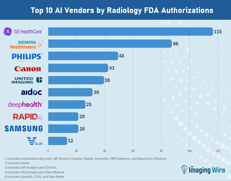
  • GE HealthCare retains the top spot as the company with the most radiology AI authorizations, at 115 (including recent acquisitions Bay Labs, BK Medical, Caption Health, MIM Software, icometrix, and Spectronic Medical).
  • Next is Siemens Healthineers at 86 (including Varian), then Philips at 48 (including DiA Analysis and TomTec), Canon at 41 (including Vital Images and Olea), United Imaging at 38, and Aidoc at 30. 

As always, it’s worth noting that the FDA’s list includes not only standalone software applications, but also imaging equipment that might have AI applications embedded into it, such as a mobile X-ray system with AI algorithms for detecting emergent conditions. 

  • Also, the agency noted that it is exploring ways to identify and tag AI-based devices that use foundation models and large language models. The FDA has yet to approve an LLM-based medical device.

The Takeaway

The new numbers indicate that radiology’s dominance of medical AI continues. But they also show that the FDA has returned to a regular twice-yearly cadence of updating its list of AI-enabled medical devices after a break of nearly a year – news that’s welcome to AI developers.

An All-in-One Radiology Platform Built for the AI Era

Early in the COVID pandemic, software engineer Shiva Suri found himself working from home alongside his radiologist mother in his parents’ basement. What he saw would lead him to build New Lantern, an AI-native platform set to disrupt the legacy radiology software market.

Suri witnessed his “world-class radiologist” mom wasting far too much time switching between five different PACS platforms and repeating the same cumbersome reporting processes with each case.

“I thought a radiologist’s job was supposed to be playing Sherlock Holmes in images,” Suri recalls, “not constantly mouse-clicking all over their PACS and tab-dictating endlessly in their reporting software.”

That imperfect workflow is an unfortunate reality for today’s radiologists, who’ve seen their processes become more tedious, while their caseloads grow in both volume and complexity.

Rads Don’t Need Another Widget

Suri’s time spent working from home became the foundation for New Lantern’s bold mission:  keep radiologists’ eyes on their images and let AI do the rest. 

  • That mission evolved over time, as Suri’s first attempt at solving radiology’s efficiency problem was a widget to automate report impressions.
  • Radiologists loved it, but… each wave of praise came with requests for more automation, leading Suri to realize that radiology’s problems weren’t going to be solved with another widget. The solution had to be fundamentally different.

The Time Is Right for an All-in-One Solution

Developing radiology’s go-to reading and reporting platform had to start with radiologists’ dream state, with their eyes on the viewer, reading image after image. 

  • It had to be based on the understanding that this dream can’t be achieved while radiologists are navigating a loosely integrated software stack.
  • The good news is, now is the perfect time to solve radiology’s software problem. The radiologist shortage and surging imaging volumes are finally driving radiology practices to look for new tech partners, and the emergence of generative AI is allowing startups to gain traction in segments that have long been dominated by entrenched legacy players. 

Enter New Lantern Curie

This perfectly timed mix of tech and market readiness set the stage for Curie, New Lantern’s all-in-one platform that combines a smart worklist, cloud PACS viewer, and AI reporter to produce AI-automated radiology report drafts.

Radiology report automation is no small task, and there’s a lot that goes into Curie’s ability to automate over 75% of non-diagnostic radiology work…

  • Streamlined Dictation – Radiologists free-dictate positive findings (no punctuation or commands), and the AI weaves them into complete sentences, generates guideline-based impressions (calculating BI-RADS, etc.), and flags errors.
  • No Tech Translations – Curie uses OCR technology to decipher technologist worksheets, applies clinical context via an LLM, and intelligently places data in the right report sections.
  • Remove Repetition – Radiologists no longer need to dictate measurements or enter prior dates. Curie handles these and a long list of other duplicative tasks for them.

The Numbers Tell the Story

All of these automations really add up, giving radiologists over 100 minutes back per shift, so they can get more done and get their lives back.

Here’s one real-world example presented at SIIM 2025 of a radiologist’s process for reading a pulmonary embolism CTA chest exam, before and after Curie…

  • Words dictated — 205 vs. 57
  • Punctuation marks & commands — 19 vs. 0
  • Fields navigated — 32 vs. 1
  • Metadata entries — 8 vs. 0 

In this example, Curie produced the same complete, accurate report with 72% fewer dictated words and 97% less navigation through dictation fields and hanging protocol changes. That’s one type of “AI taking radiologists’ jobs” that just about every radiologist would welcome.

The Takeaway

As imaging volumes surge and antiquated platforms push radiologists to the breaking point, New Lantern Curie offers them a way to work like it’s 2025 instead of 2005 – automating the fragmentation and duplication out of their days so world-class radiologists like Shiva Suri’s mom can focus on what they do best: reading images.

Learn more about New Lantern and its all-in-one approach to radiology workflow in this Imaging Wire Show video interview

Could States Take Over AI Regulation from the FDA?

Could states take over AI regulation from the FDA as a possible solution to the growing workforce shortage in radiology? It may seem like a wild idea at first, but it’s a question proposed in a special edition of Academic Radiology focusing on radiology and the law. 

Healthcare’s workforce shortage is no secret, and in radiology it’s manifested itself with tight supplies of both radiologists and radiologic technologists. 

  • AI has been touted as a potential solution to lighten the workload, such as by triaging images mostly likely to be normal from requiring immediate radiologist review. 

And autonomous AI – algorithms that operate without human oversight – are already nibbling at radiology’s fringes, with at least one company claiming its solution can produce full radiology reports without human intervention.

  • But the FDA is notoriously conservative when it comes to authorizing new technologies, and AI is no exception. So what’s to stop a state facing a severe radiologist shortage from adopting autonomous AI on its own to help out? 

The new article reviews the legal landscape behind both constitutional and state law, finding examples in which some states have successfully defied federal regulation – such as by legalizing marijuana use – if the issue has broad public support. 

But the authors eventually answer their own question in the negative, stating that it’s not likely states will usurp the FDA’s role regulating AI because…

  • The U.S. Constitution’s Supremacy and Commerce clauses ensure federal law will always supersede state law.
  • If AI made an error, malpractice regulation would be murky given a lack of legal precedent at the state level. 
  • Teleradiologists could opt out of providing care to a state if AI regulations were too burdensome – which could exacerbate the workforce crisis. 

The Takeaway

Ultimately, it’s not likely states will take over AI regulation from the FDA, even if the healthcare workforce shortage worsens significantly. But the Academic Radiology article is an interesting thought experiment that – in an environment in which U.S. healthcare policies have already been turned upside down – may not be so unthinkable after all. 

AI Predicts Radiology Workload

AI is touted as a tool that can help radiologists lighten their workload. But what if you could use AI to predict when you’ll need help the most? Researchers in Academic Radiology tried that with an AI algorithm that predicted radiology workload based on three key factors. 

Imaging practices are facing pressure from a variety of forces that include rising imaging volume and workforce shortages, with one recent study documenting a sharp workload increase over the past 10 years.

  • Many industry observers believe AI can assist radiologists in reaching faster diagnoses, or by removing studies most likely to be normal from the worklist based on AI analysis. 

But researchers and vendors are also developing AI algorithms for operational use – arguably where radiology practices need the most help.

  • AI can predict equipment utilization, or even create a virtual twin of a radiology facility where administrators can adjust various factors like staffing to visualize their impact on operations.

In the new study, researchers from Mass General Brigham Hospital developed six machine learning algorithms based on a year of imaging exam volumes from two academic medical centers.

The group entered 707 features into the models, but ultimately settled on three main operational factors that best predicted the next weekday’s imaging workload, in particular for outpatient exams…

  • The current number of unread exams.
  • The number of exams scheduled to be performed after 5 p.m.
  • The number of exams scheduled to be performed the next day.

The algorithm’s predictions were put into clinical use with a Tableau dashboard that pulled data from 5 p.m. to 7 a.m. the following day, computed workload predictions, and output its forecast in an online interface they called “BusyBot.”

  • But if you’re only analyzing three factors, do you really need AI to predict the next day’s workload? 

The authors answered this question by comparing the best-performing AI model to estimates made by radiologists from just looking at EHR data. 

  • Humans either underestimated or overestimated the next day’s volume compared to actual numbers, leading the authors to conclude that AI did a better job of calculating dynamics and weighting variables to produce accurate estimates.

The Takeaway

Using AI to predict the next day’s radiology workload is an intriguing twist on the argument that AI can help make radiologists more efficient. Better yet, this use case helps imagers without requiring them to change the way they work. What’s not to like?

AI and Legal Liability in Radiology

What impact will artificial intelligence have on the legal liability of the radiologists who use it? A new study in NEJM AI suggests that medical malpractice juries may pass harsher judgment on radiologists when they make mistakes that disagree with AI findings.

AI is viewed as a technology that can save radiologists time while also helping them make more accurate diagnoses.

  • But there’s a dark side to AI as well – what happens when AI findings aren’t correct, or when radiologists disagree with AI only to discover it was right all along?

In the new study, a research team led by Michael Bernstein, PhD, of Brown University queried 1.3k U.S. adults on their attitudes toward radiologists’ legal liability in two clinical use cases for AI – identifying brain bleeds and detecting lung cancers.

  • Participants were asked if they felt radiologists met their duty of care to patients across different scenarios, such as whether the AI and the radiologist agreed or disagreed on the original diagnosis. 

Responses were compared to a “no AI” control scenario in which respondents assessed legal liability if radiologists hadn’t used AI at all, with researchers finding …

  • If radiologists disagreed with AI, more respondents found radiologists liable …
    • Brain bleeds: 73% found radiologist liable (vs. 50% with no AI)
    • Lung cancer: 79% found radiologist liable (vs. 64% with no AI)
  • If both radiologists and AI missed the diagnosis, there was no statistically significant difference …
    • Brain bleeds: (50% vs. 56% with no AI, p=0.33)
    • Lung cancer: (64% vs. 65% with no AI, p=0.77)
  • Respondents were less likely to side with plaintiffs when given information about standard AI error rates …
    • When AI agreed with the radiologist diagnosis:
      • Brain bleeds: (73% plaintiff agreement fell to 49%)
      • Lung cancer: (79% fell to 73%)
    • When AI disagreed with the radiologist diagnosis:
      • Brain bleeds: (50% plaintiff agreement fell to 34%)
      • Lung cancer: (64% fell to 56%)

The Takeaway

The new study offers a fascinating look at AI’s future in radiology from a medico-legal perspective. But there’s one question the researchers didn’t address: If AI-supported image interpretation eventually becomes the standard of care, will radiologists be found liable for not using it at all? Stay tuned. 

Get every issue of The Imaging Wire, delivered right to your inbox.