Cochrane Pivots on Prostate Screening

Prostate cancer screening is getting new support from an unlikely source – the Cochrane group, which historically has been skeptical of population-based screening. Cochrane researchers last week published a new report supporting prostate screening, a sharp change from the group’s previous guidance. 

Prostate cancer screening hasn’t achieved the generally accepted status of other cancer screening tests like breast, cervical, colorectal, and lung. 

  • One of the main sticking points has been overdiagnosis. Prostate cancer can often be slow-growing, and many men live for years with prostate disease before dying of other causes.

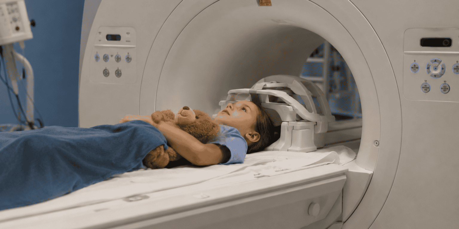
But that dynamic has been changing in recent years, in large measure due to the ability of MRI to differentiate aggressive prostate cancer from more indolent disease. 

  • Clinicians are incorporating MRI into prostate screening protocols, using it to determine which men with elevated PSA levels should be biopsied and which ones can be followed with active surveillance. 

For its part, Cochrane is an international non-profit research consortium that periodically analyzes the peer-reviewed evidence behind new medical exams and technologies. 

  • But Cochrane’s work has occasionally been controversial: The group last month published a negative review of Alzheimer’s drugs that included treatments that never made it to market. Also, a Cochrane research center in Denmark for years was one of the most vociferous opponents of mammography screening. 

So that’s why last week’s statement on prostate screening is so surprising, especially given that Cochrane’s 2013 review found no evidence to support the claim that screening reduced prostate cancer mortality. 

In the new review, Cochrane analyzed data from six clinical trials in Europe and North America that included 800k men, finding that screening with PSA blood tests…

  • Detected 30% more prostate cancers overall, most at an early stage. 
  • Reduced the relative risk of a metastatic prostate cancer diagnosis by 35%.
  • Reduced prostate cancer mortality by 2 deaths for every 1k men screened (for comparison, mammography’s benefit is estimated to be 6-8 deaths). 
  • For every 1-2 deaths prevented, 36 additional cancers were diagnosed – a possible sign of overdiagnosis. 

What changed? Cochrane researchers said that we now have longer-term data that makes it easier to detect screening’s subtle mortality benefit.

  • They also cited the success of technologies like MRI in reducing unnecessary biopsies – and the harms of overdiagnosis.

The Takeaway

Last week’s news suggests that the ground is shifting under prostate cancer screening in favor of broader use of the exam, potentially with MRI follow-ups. If you can convince a screening-skeptical group like Cochrane of prostate screening’s value, you can convince anyone. 
AI for PE Detection: ‘Selective but Meaningful’

AI made a “selective but meaningful” contribution to radiologist interpretations of CT pulmonary angiography scans for pulmonary embolism. The study, published in Radiology: Artificial Intelligence, offers valuable insights into real-world implementation of AI on a large scale. 

One of the major criticisms of AI is that algorithms used in real-world clinical situations don’t perform as well as they do in the controlled environments that vendors use to acquire data for regulatory submissions.

  • AI performance can drop off as much as 20 to 30 percentage points for important metrics like sensitivity and specificity. 

The new study sought to investigate this phenomenon by analyzing a real-world implementation of Aidoc’s AI algorithm for PE detection. 

  • Researchers assessed the algorithm’s performance for analyzing CTPA exams across a variety of clinical environments in an integrated health network, including the emergency department and inpatient and outpatient settings. 

Scans of 29.5k patients acquired from 2021 to 2023 were included. AI analyzed images in real time, after which exams were interpreted by radiologists who knew the AI findings. Researchers found…

  • Radiologists using AI had higher sensitivity than the algorithm on its own (99% vs. 85%).
  • Specificity was more or less the same (99.8% vs. 99.5%).
  • Agreement between radiologists and AI was high (98%).
  • Agreement was higher when AI assessed cases as negative rather than positive (98% vs. 94%).
  • Radiologists disagreed with AI in 2.2% of cases. The final determination by a panel of expert thoracic radiologists strongly favored radiologists (89%).
  • Of the 3.3k cases positive for PE, 0.81% were detected only by AI – or 26 cases.

In analyzing the results, the researchers characterized AI’s contribution as “selective but meaningful.”

  • AI-positive results meant scans might require more scrutiny from radiologists, while an AI-negative call might be supportive – but not definitive – for negative PE.

The Takeaway

The new study of AI for PE detection is a fascinating look at real-world AI deployment. While the sensitivity, specificity, and agreement numbers are interesting, what draws our attention is the 26 PE cases caught only by AI over 18 months of use. That boils down to 26 patients whose clinical condition wasn’t missed, and 26 potential malpractice lawsuits that were never filed.

Pediatric MRI Safety Surveyed

This past year has seen a renewed focus on MRI safety after a fatal accident in New York in 2025. Most of the attention has been on adult MRI, but what about kids? A new analysis in JACR finds that pediatric MRI accidents are fortunately rare, but occur often enough that continued vigilance is warranted. 

Pediatric MRI poses particular safety challenges to imaging facilities. Caregivers are often in the scanning room to comfort children, and sedation is frequently required to keep kids still during exams. 

  • But pediatric MRI safety hasn’t been studied as extensively as it has in adults, so researchers from five U.S. children’s hospitals reviewed MRI safety events that occurred at their facilities from 2017 to 2022.

Researchers focused on reported events that occurred within Zone IV, the area under the ACR’s four-zone safety model that includes the scanner room. They found…

  • A total of 146 safety events occurred in Zone IV out of 541k pediatric exams, for an event rate of 0.027%. 
  • An average of 4.9 events per year occurred at each site, or 3.3 events per 100k exams.
  • Event types involved projectiles (30%), burn/thermal injuries (13%), and implants (10%).
  • 78 events (53%) directly involved patients.
  • Ten events (6.8%) were classified as serious.

Frequent causes of events included medical equipment and supplies (anesthesia equipment and monitors, stethoscopes, and needles) and personal items like phones and badges.

  • Implanted devices like cochlear implants represent a growing challenge, as 20%-30% of children getting MRI scans have them, and safety events occurred despite sites following manufacturers’ guidelines. 

Why did the safety events happen? Study authors found that MRI safety protocols weren’t followed in 60% of events.

  • Lack of protocol adherence is a common refrain in MRI accidents that have involved adults, illustrating that all the guidelines and rules in the world won’t help if they aren’t followed to the letter.

The Takeaway

The new study on pediatric MRI safety highlights the fact that children shouldn’t just be treated like little adults when it comes to safe scanning procedures. The research offers a benchmark against which pediatric imaging facilities can measure themselves, while also offering additional guidance on mistakes to avoid when scanning kids.

Imaging Predicts Disease by Analyzing Tissue Composition

Two new studies published this week highlight the exciting potential of medical imaging scans to guide preventive health by analyzing the composition of tissues like muscle and fat. The research suggests that imaging can predict a person’s risk of serious disease years before symptoms occur.

Previous research has shown that the composition of various types of body tissue can serve as biomarkers for future health problems, particularly cardiovascular disease and metabolic issues like diabetes.

  • Advances in AI analysis are driving much of the new understanding, as increasingly powerful deep learning algorithms are emerging that can analyze massive quantities of imaging data and compare tissue composition to databases of normal scans.

The new studies – both published in Radiology – test this concept on a large scale. In the first, researchers in Germany reviewed MRI data from over 66k people who got whole-body MRI scans as part of large population health studies (UK Biobank and German National Cohort, or NAKO). 

  • They used deep learning algorithms to create z-scores of tissue composition metrics and correlated those measurements to the risk of developing conditions like diabetes and cardiac events, as well as all-cause mortality.

Researchers found…

  • High visceral adipose tissue indicated higher risk of incident diabetes (HR = 2.26).
  • High intramuscular adipose tissue was connected to major adverse cardiovascular events (HR = 1.54).
  • Low skeletal muscle was linked to all-cause mortality (HR = 1.44).

In the second study, researchers focused on 11.3k participants in the NAKO study who got whole-body 3T MRI scans.

  • Researchers used software to analyze two tissue composition metrics, paraspinal intermuscular adipose tissue (IMAT) and lean muscle mass (LMM), which only recently have been connected to metabolic dysfunction.

The authors found…

  • Increased IMAT was associated with higher risk of hypertension and atherogenic dyslipidemia, a lipid imbalance associated with metabolic dysfunction and diabetes risk (HR = 1.67 and 1.82, respectively).
  • Higher LMM was a marker for better health in men, and was linked to lower odds of hypertension and atherogenic dyslipidemia (HR = 0.34 and 0.49, respectively). 

The Takeaway

The new studies build on previous research to show how the combination of imaging-derived biomarkers and AI-based analysis can pinpoint currently healthy people who might be at higher risk of future disease. The implications are exciting for anyone who believes radiology can play a greater role in guiding population health.

Radiologist Tapped As Surgeon General

Could America’s next top doctor be a radiologist? The radiology world – and the rest of U.S. healthcare – was stunned late last week when the Trump Administration nominated radiologist Nicole Saphier, MD, to be surgeon general, replacing previous nominee Casey Means, MD.

If confirmed, Saphier’s nomination would be the first time a board-certified radiologist has held the position, which typically goes to physicians with experience in public health rather than medical specialists.

  • Trump nominated Means for the position in May 2025, but the nomination languished over concerns about Means’ experience, her lapsed medical license, and her tepid support for vaccines.

On the other hand, Saphier is an actively practicing radiologist who serves as director of breast imaging at Memorial Sloan Kettering Monmouth in New Jersey.

  • She’s also been a frequent contributor to Fox News, where she appeared on the conservative network’s “Fox & Friends” morning show as an expert on public health policy. 

Saphier was born and raised in Arizona, where she completed her radiology residency and was involved in efforts in 2014 to pass breast density notification legislation in the state. 

  • She moved to New Jersey later that year and worked in a private-practice breast imaging center before taking the position she currently holds at MSK Monmouth. 

Saphier has always been active on social media (her X account has 364.4k followers), due to her belief that radiologists should be more visible to patients.  

  • It was that presence that initially drew the attention of Fox News producers, and Saphier began appearing on the network in 2016 to comment on public policy issues (her involvement with Fox ended with the nomination announcement).

Saphier’s nomination is already drawing critics who are combing over her history of statements on vaccines and the government response to the COVID-19 pandemic. 

  • In general, Saphier has expressed skepticism about government involvement in healthcare, but most of her beliefs fall within the mainstream of U.S. public health policy, which should bode well for her nomination.

ACR issued a statement supporting Saphier’s nomination, noting her work with the group on several public policy issues and observing that if confirmed, “Saphier would be the highest-ranking radiologist ever in government service.”

The Takeaway

Politics aside, Saphier’s ascension as surgeon general could have huge benefits for radiology in general and breast imaging in particular. Saphier has consistently supported mammography screening and issues like breast density awareness, and should her nomination succeed, radiology would find itself with an ally at the highest levels of the U.S. government. 

Two Views on Lung Screening’s Progress

Two new research studies published this week offer slightly conflicting views on the progress of CT lung cancer screening. One study saw the screening adherence rate rising to nearly 25%, while another revealed that screening might not be as effective in the real world as it was in the landmark National Lung Screening Trial. 

Low-dose CT lung cancer screening was approved for Medicare reimbursement in 2015 following promising NLST results, but the exam was slow to catch on.

  • That’s begun to change as U.S. physicians identify how to get eligible high-risk people into screening, while other countries are launching organized population-based screening programs, Germany being the most recent.

In the first study this week, published in JAMA Internal Medicine, researchers calculated the most recent uptake rate for LDCT lung cancer screening.

  • Data from 26.1k people from the BRFSS survey in 2024 were analyzed and compared to 2022 data.

The rate of up-to-date LDCT screening prevalence in 2024…

  • Rose six percentage points (24% vs. 18%).
  • Differed by just 0.5 percentage points between men and women.
  • Was highest and rose the most for people ages 65-69 (33%, increasing by 9 percentage points).
  • But still lagged screening rates for other cancers like breast (80%), cervical (75%), and colorectal (67%).

This week’s second study was published in JAMA Network Open and offers additional context, finding that real-world lung screening may not reduce all-cause mortality by as much as what was seen in the NLST.

  • Researchers analyzed screening’s effectiveness in a program run by the U.S. Veterans Health Administration, which has one of the more successful lung screening programs in the country.

They compared five-year all-cause mortality rates in the VA program for 732 people who were screened, finding that, compared to NLST…

  • Mortality rates were 2.5X higher in the VA population (24% vs. 9.7%).
  • VA participants had 3X the mortality risk (HR = 2.98).

VA researchers noted that while their population met the same screening eligibility criteria as NLST, the VA patients were sicker and probably less likely to see as much long-term benefit from LDCT screening.

The Takeaway

This week’s studies are a window into the complex changes underway in CT lung cancer screening. While screening’s rising participation rate is good news, the mortality findings show that estimating screening’s effectiveness is more complex than simply overlaying NLST results on real-world populations. 

Mammography AI Improves Breast Screening

Radiologists using a commercially available mammography AI algorithm saw improved diagnostic performance in breast cancer screening, mainly due to better specificity. The study adds to a growing body of research supporting mammography AI.

Mammography screening has been one of the most promising use cases for AI, and recent randomized controlled trials have demonstrated that AI can both improve diagnostic accuracy and speed up workflows. 

  • But RCTs are usually performed under highly controlled conditions in high-income Western countries, and the results might not be generalizable to other countries around the world. 

In the new study in Academic Radiology, researchers in Singapore tested Lunit’s Insight MMG algorithm in a retrospective review of a dataset of 302 digital mammograms that was enriched with 89 breast cancers.

  • Researchers noted that many countries have a high breast cancer incidence-to-mortality ratio due to limitations in population-based screening programs, and AI potentially could help. 

The authors focused on AI’s ability to improve the diagnostic performance of nine breast radiologists from four countries in Asia and North Africa who interpreted the mammograms, finding that AI assistance…

  • Improved radiologist accuracy as measured by AUC (from 0.799 to 0.851).
  • Generated a big jump in specificity (from 77% to 88%). 
  • And significantly reduced per-case image interpretation times (from 122 to 83 seconds per case).
  • Without changing sensitivity at a statistically significant level (83% vs. 82%, p = 0.73).

There were some subtle differences in the current study’s findings relative to previous research, some of which were the result of using a cancer-enriched dataset rather than a screening population as would be the case in an RCT.

  • The specificity improvement with AI would reduce unnecessary recalls in a population-based screening program and make mammography more cost-effective – an important consideration in countries with constrained public health budgets.

The Takeaway

The new study doesn’t have the statistical heft of a large, randomized controlled trial, but it still adds to the body of knowledge supporting AI for mammography, especially at facilities that haven’t been party to the large-scale RCTs.

Physician Burnout Eases – But Remains High in Radiology

Physician burnout rates continue to drop after peaking during the COVID-19 pandemic, but radiology remains one of the medical specialties with the worst burnout problem. That’s according to a new AMA survey that underscores a continuing trend toward improved physician satisfaction.

Physician burnout has been a cause of concern for the past decade. Burnout rates accelerated dramatically during the COVID-19 pandemic as doctors struggled with long hours and crushing patient volume. 

The new AMA survey confirms the trend. The organization surveyed 19k U.S. physicians throughout 2025, finding…

  • An overall physician burnout rate of 42% in 2025, down compared to 2024 (43%) and 2023 (48%), and sharply down from the peak in 2021 (63%).
  • Physician job satisfaction rose to 77%, stable compared to 2024 (77%) and up compared to 2023 (72%) and 2022 (68%).
  • The burnout rate in radiology was 45%, making it the fifth-worst among specialties after emergency medicine (50%), urological surgery (50%), hematology/oncology (49%), and OB/GYN (46%). 

How well do the AMA’s numbers reflect radiology’s reality?

  • Unfortunately, the AMA hasn’t included radiology-specific numbers in previous years of its survey, making it hard to directly compare year-over-year changes.

But in a recent thread on radiology bulletin board RadHQ.net, members anecdotally reported that burnout is becoming less prominent for radiologists, for several reasons…

  • Higher compensation that makes radiologists feel more valued. 
  • More flexible arrangements – like teleradiology – that support working from home or more flexible shifts.
  • Increased job mobility due to the radiologist shortage, with the flexibility to leave toxic practices for new ones.

The Takeaway

So which is it – is radiology burnout higher than other specialties, or is it declining? Ironically, both statements could be true – while radiologist burnout remains high relative to other physicians, at least it’s trending in the right direction.

Radiologist Training Stagnates

The number of new radiologists being trained in the U.S. has largely stagnated, even as concerns grow over workforce shortages. A new analysis in JACR underscores the challenges facing efforts to train more radiologists to meet the rising volume of imaging exams.

The growing imbalance between the radiologist workforce and surging imaging volume has been generating headlines as frontline radiologists struggle with overwork and burnout. 

  • And a recent effort to boost the number of U.S. physicians by adding federal funding for more resident training slots has overlooked radiology, which is getting few of the new positions being offered.

In the new study, researchers analyzed the number of U.S. resident positions in diagnostic and interventional radiology using data from the NRMP, ACGME, and other sources, finding…

  • The number of radiology residency positions grew 33% from 2010 to 2025 (from 1,090 to 1,449), while total medical residency positions grew 69%.
  • The total number of radiology residents rose 23% from 2010 to 2024 (from 4,584 to 5,630). 
  • But the number of practicing radiologists only grew 12% from 2010 to 2022 (from 34.3k to 38.3k). 
  • And the ratio of radiologists per 100k population was stagnant (from 11.1 to 11.5 radiologists).

It’s this last number that’s key to the analysis. Over the last 15 years, the ratio of radiologists to the U.S. population has barely changed, even as imaging exams become more complex.

  • And even more importantly, the Baby Boom generation has aged and now requires more healthcare services per capita – including imaging – relative to when they were younger (a previous study found the average radiologist workload doubled from 2008 to 2018). 

The researchers conclude that the radiologist training pipeline “has barely kept pace” with the increasing U.S. population, and when coupled with growing complexity and per capita imaging use, this raises questions about the sustainability of increasing imaging utilization. 

  • They recommend more federal support for radiologist training positions, perhaps through Medicare programs that specifically target medical imaging.

The Takeaway

The new study on stagnant radiologist training gives ammunition to radiology advocates who are lobbying for more federal funding to expand radiology residency slots. The question is whether anyone is listening.

Radiologist Salaries Grow 9%

It’s not quite double-digit growth, but it’s pretty close. The latest physician salary report from Medscape reveals that radiologist salaries grew 9% in 2025, with medical imaging seeing the third-highest growth rate among the specialties tracked. 

The new Medscape report found that radiologists had an average annual salary of $571k in 2025, up 9% compared to $520k in 2024 and $498k in 2023.

  • Radiology ranked #3 among the 29 physician specialties tracked by the survey, trailing only orthopedic physicians and surgeons ($611k) and cardiologists ($575k). 

Radiology’s numbers mark the second straight year of strong salary growth for the specialty, a welcome trend given anemic growth in 2023, when salaries rose only 3.1% – not enough to keep ahead of inflation.

  • Indeed, industry experts analyzing the new Medscape numbers called the figures a “return to normalization” for physician pay gains. 

Medscape reported a generally “positive vibe” to the survey compared to a year earlier, with more respondents saying they felt fairly paid (53% vs. 48%), and eight specialties reporting average annual compensation over $500k. Other important findings included…

  • Technology-driven efficiency appears to be helping physician productivity, enabling doctors to see more patients and generate more work-related relative value units.
  • Rising demand for services in orthopedics, radiology, and cardiology is driving pay growth, especially given the static supply of physicians.
  • The new data – and improving physician sentiment – could indicate that medicine is climbing out of the trough of doctor burnout that bottomed out during the COVID-19 pandemic. 
  • The gender gap between salaries for male and female physicians was $102k, worsening slightly compared to last year (31% vs. 29%). 
  • The average workweek shortened slightly (49 vs. 50 hours).

The Takeaway
Medscape’s new salary survey findings are positive news for radiologists – and indeed all physicians – and are a welcome departure from the days of burnout and stagnant wage growth. So far, predictions that AI will take jobs from radiologists remain far in the future.

Get every issue of The Imaging Wire, delivered right to your inbox.