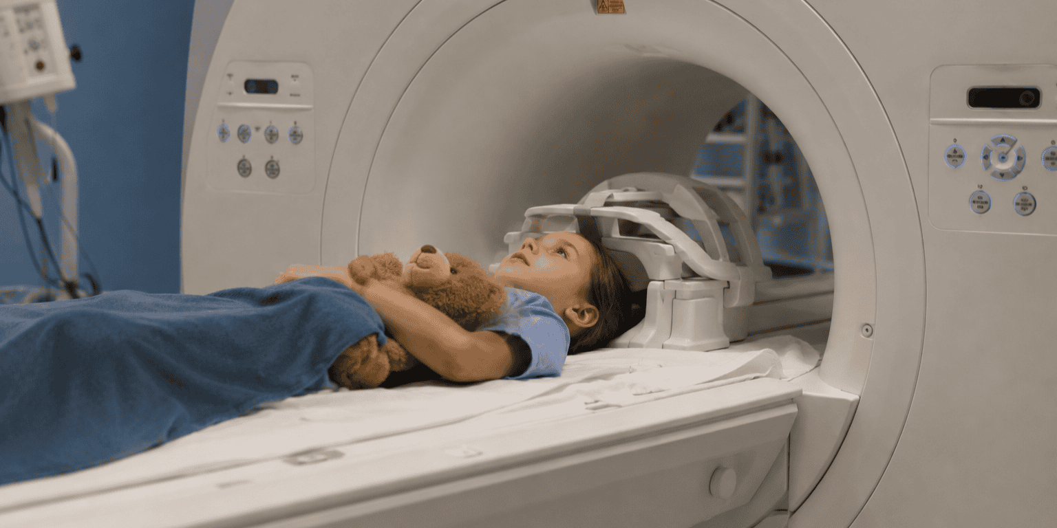
Molecular MRI Adds Certainty to Cancer Diagnosis

MRI has become an important tool in the detection, diagnosis, and treatment planning for many cancers, especially solid tumors. However, up until now, a lack of specificity has held back the full potential of MRI.  

While MRI is very good at identifying areas of interest, factors such as infection, benign tumors, post-traumatic areas, and inflammation can all increase vascularity and, therefore, enhancement of contrast and signal changes.  

  • As a result, MRI has a high rate of false positives – findings that may be flagged as something of concern but that are not necessarily malignant lesions.  

This lack of accuracy results in clinical care teams performing too many confirmatory biopsies, with most being benign.

Now a novel class of molecular imaging contrast agents developed by Imagion Biosystems brings a new level of specificity to MRI. 

  • The company’s MagSense imaging agents have the potential to improve the clinical utility of the large installed base of MRI systems across the globe through improved accuracy of interpretation, avoiding biopsies of benign lesions, driving earlier intervention and improving outcomes and quality of life.

Unlike gadolinium-based agents that non-specifically enhance tissue vascularity regardless of cause, MagSense imaging agents target receptors on cancer cells.  

  • By combining magnetic nanoparticles that have high susceptibility and r2 relaxivity with cancer-specific biomarkers, molecular MRI becomes possible.

Imagion’s superparamagnetic iron oxide nanoparticles are coated with a cancer-specific targeting moiety, such as an antibody or peptide.

  • The cancer biomarker molecule causes the particles to bind to target-specific cancer cells, if present. If the lesion in question is not the target cancer, the particles do not bind.

Where the imaging agent has become attached to the tissue, the nanoparticles produce an identifiable change in MRI signal. 

  • This signal is easily detected by radiological review and can be quantitatively assessed.

Imagion has developed cancer-specific contrast imaging agents for HER2 breast cancer, prostate cancer, and ovarian cancer, and the MagSense platform can be adapted for any type of cancer for which there is a targeting moiety.  

  • Imagion is now preparing to initiate a multisite phase 2 study in the U.S. in HER2+ breast cancer patients to optimize imaging parameters and compare MagSense imaging to the standard of care.  

The Takeaway

Molecular-specific imaging agents like the MagSense technology from Imagion Biosystems create the opportunity for molecular MRI to fundamentally change how radiologists detect and monitor cancers. 

The company is publicly traded (ASX:IBX) and is looking to expand its U.S. investor base as it advances through its clinical programs. To become involved as an investigator or investor or to learn more visit their website.

Imaging Workload Jumps with Higher Use of CT, MRI

Radiology’s shift to more advanced modalities like CT and MRI is increasing the burden on radiologists to interpret more complex exams. A new study in JACR documents the trend, finding that radiologist workload for inpatient imaging has risen sharply over the last 10 years. 

Like many physicians, radiologists are feeling burned out from rising patient workload, personnel shortages, and declining reimbursement. 

  • But radiology has the added burden of being one of healthcare’s most technology-focused specialties, with new imaging modalities giving them cooler tools to work with, but at the cost of steadily increasing exam complexity.

Researchers from Brigham and Women’s Hospital have been tracking inpatient imaging utilization for the past 40 years, and the new paper provides the latest update. 

  • They calculated inpatient imaging volume at Brigham and Women’s from 2012 to 2023, during which 896k imaging exams were performed.  

Results for the study were as follows …

  • Total annual inpatient imaging volume grew 17% over 10 years (102k to 119k exams).
  • Total imaging exams per patient admission (adjusted by case mix and disease severity) fell 20% due to declines in X-ray, ultrasound, and nuclear medicine.
  • But imaging exams per patient admission grew for CT (19%) and MRI (21%).
  • Leading to growth in CT and MRI’s combined share of all radiology global RVUs (62% to 75%).
  • Hospital length of stay rose 32% (5.6 to 7.4 days), possibly due to the COVID-19 pandemic. 

What does it all mean? Basically, the number of inpatient imaging exams per patient is declining when adjusted for disease severity, but radiologists are still having to work harder because the studies are more complex. 

  • Imaging could also be shifting from the inpatient setting to outpatient centers due to reimbursement changes aimed at shifting exams to lower-cost settings than hospitals.

One big question with the new study is the degree to which the COVID-19 pandemic skewed the results compared with previous years. 

  • The pandemic may have spurred more use of CT, especially given its value in providing a definitive diagnosis of SARS-CoV-2 infection. 

The Takeaway

If you feel like you’re working harder than ever, the new findings show that you’re not crazy. And given radiology’s breakneck pace of innovation, it’s not likely the trends revealed in the new study will let up any time soon.

MRI in Paradise – News from ISMRM 2025

The global MRI community this week traveled to paradise to convene its annual meeting of the International Society for Magnetic Resonance in Medicine. If you were one of the lucky ones to be in attendance in Honolulu, Hawaii for ISMRM 2025, you were treated to some of the latest news in radiology’s most powerful modality. 

As has been the case at other radiology meetings, AI took center stage in Honolulu. 

  • AI has multiple use cases in MRI, from helping radiologists interpret images more efficiently to accelerating scans and upscaling lower-field images to resemble high-field exams.

Just a few of the news highlights from ISMRM 2025 are below …

  • Using AI to interpret prostate MRI reduced reading times by 48% (250 to 120 seconds) while improving the diagnostic performance of both experienced and less experienced radiologists. 
  • AI of thyroid T2-weighted neck MRI scans demonstrated good accuracy (87%) for nodules larger than 1 cm, indicating a possible role for screening and monitoring.
  • Researchers presented progress in creating brain charts of white matter based on MRI scans of 24k cognitively healthy people that can be used to track normal and abnormal brain development.
  • Brain MRI showed that lower brain volumes in people with coronary artery disease were associated with worse aerobic fitness and higher BMI, revealing a link between cardiovascular and brain health. 
  • Chinese researchers showed their work on PMEEN, a multimodality brain scanner that combines PET, MRI, EEG, eye-tracking, and functional near-infrared spectroscopy. 
  • A Spanish team demonstrated research on a low-field PET/MRI scanner with focused ultrasound capability for therapeutic applications.
  • AI could be used during abbreviated breast MRI screening scans to convert women mid-exam to a full MRI protocol if abnormalities are detected.
  • 7T MRI was used to detect iron deposits in the brain, which could be a marker for Alzheimer’s disease.
  • MRI with an ultrashort echo time protocol could be an alternative to CT for following up lung nodules.
  • Researchers presented a deep learning-based approach to generating synthetic contrast-like MR images without gadolinium. 
  • MGH researchers showed progress in developing a 136mT portable MRI scanner for bedside brain scanning of preterm neonates.

The Takeaway

The rapid proliferation of news about AI-based MRI at ISMRM 2025 suggests its own vision of paradise – a world in which MRI can be deployed more widely than ever before, where radiologists with AI assistance detect disease in many cases before symptoms even occur. We can only dream.

MRI Recon Gets Real with AI-Driven Protocols

AI-based data reconstruction for MRI scans took a step forward this week with studies showing how to generate 3T-like images from ultralow-field scanners, and improve scanner efficiency by cutting energy consumption.

MRI is radiology’s premier modality, but MRI scanners are cumbersome to install and expensive to operate. 

  • Ultralow-field scanners could help but some believe they lack the image quality for some clinical applications. 

Enter AI-based image reconstruction. Deep learning protocols are being developed for a wide range of imaging modalities, from PET to CT to MRI. 

  • These algorithms take images acquired with lower-quality input data – be it less CT radiation dose or lower MRI field strength – and upscale them to resemble full-fidelity images.

This trend is illustrated by research published this week in Radiology in which researchers tested a generative adversarial network algorithm called LowGAN for reconstructing data acquired on Hyperfine’s Swoop 0.064T portable ultralow-field MRI scanner.

  • Their goal was to enable Swoop to generate images resembling those acquired on a 3T system. 

After training LowGAN on paired 3T and 0.064T images, they tested the algorithm in 50 patients with multiple sclerosis and further validated it with a separate 13-patient cohort. They then judged LowGAN against several measures of MR image quality, finding that it …

  • Showed the biggest improvement on synthetic FLAIR and T1 images.
  • Improved conspicuity of white matter lesions, without introducing false lesions.
  • Increased consistency of cortical and subcortical volume measurements with 3T images.
  • But was unable to reveal brain lesions that were missed in the original low-field scans. 

AI-based data reconstruction also has environmental implications. Medical imaging is a major contributor to greenhouse gas emissions, and anyone who’s managed an MRI operation knows how much energy these massive scanners consume. 

  • A second paper published this week in Radiology described how MRI acceleration – scans acquired at a faster speed and then reconstructed for better image quality – reduced energy use, lowering carbon emissions while boosting imaging capacity. 

Researchers tried three techniques for speeding MRI acquisition – parallel acceleration, simultaneous multi-slice, and a deep learning algorithm. 

  • All three reduced energy consumption 21% to 65% and increased daily capacity by one to seven scanning slots, with deep learning showing the biggest effect.

The Takeaway

The new papers demonstrate an exciting future in which less powerful data acquisition technologies can be upscaled with AI to produce images that more closely resemble state-of-the-art scanning. The benefits will be enjoyed by both patients and the planet.

AI Helps Radiologists Read Prostate MRI

MRI is changing how prostate cancer is detected, diagnosed, and followed up. But even a technology as powerful as MRI could use a little help, as evidenced by a new study in Radiology showing that a commercially available AI algorithm could help radiologists diagnose clinically significant prostate cancer. 

Workup of suspicious prostate lesions is being reshaped by MRI in meaningful ways.

  • For example, MRI-guided biopsy is replacing systemic prostate biopsy without guidance, especially for patients with low to intermediate risk of prostate cancer. 

But prostate MRI isn’t perfect – yet. Radiologist performance can vary due to differences in experience, as well as variations in MRI acquisitions, tumor location, and cancer prevalence. Could AI help even out these variations? 

  • To find out, researchers from South Korea tested Siemens Healthineers’ syngo.via Prostate MR algorithm in 205 patients suspected of prostate cancer who were scheduled for biopsy based on clinical information (including previous MRI scans).

The AI algorithm’s performance was compared to that of experienced radiologists, and researchers also estimated its impact on radiologist interpretation if used as a reading aid, finding that for clinically significant prostate cancer… 

  • AI had lower sensitivity versus radiologists (80% vs. 93%).
  • But higher positive predictive value (58% vs. 48%).
  • Adding AI to radiologists’ interpretation more than doubled specificity (44% vs. 21%).
  • There were no cancer cases among lesions rated by both the algorithm and radiologists as not likely to be cancer (PI-RADS 1 or 2).

AI’s higher PPV indicates that it could help reduce unnecessary prostate biopsies, while also detecting clinically significant cancer that might have been missed by radiologists.  

The Takeaway

The new findings echo previous studies that demonstrate the value of AI for MRI of prostate cancer, but differ in that they investigate a commercially available algorithm – indicating that tools for better prostate MRI are becoming accessible to radiologists. 

AI Enables Single-Click Cardiac MRI

Cardiac MRI is one of the most powerful imaging tools for assessing heart function, but it’s difficult and time-consuming to perform. Could automated AI planning offer a solution? A new research paper shows how AI-based software can speed up cardiac MRI workflow

Cardiac MRI has a variety of useful clinical applications, generating high-resolution images for tissue characterization and functional assessment without the ionizing radiation of angiography or CT.

  • But cardiac MR also requires highly trained MR technologists to perform complex tasks like finding reference cardiac planes, adjusting parameters for every sequence, and interacting with patients – all challenges in today’s era of workforce shortages. 

Cardiac MRI’s complexity also increases the number of clicks required by technologists to plan exams. 

  • This can introduce scan errors and produces inter-operator variability between exams. 

Fortunately, vendors are developing AI-based software that automates cardiac MR planning – in this case, Siemens Healthineers’ myExam Cardiac Assist and AI Cardiac Scan Companion. 

  • The solution enables single-click cardiac MR planning with a pre-defined protocol that includes auto-positioning to identify the center of the heart and shift the scanner table to isocenter, as well as positioning localizers to perform auto-align without manual intervention. 

How well does it work in the real world? Researchers tested the AI software against conventional manual cardiac MR exam planning in 82 patients from August 2023 to February 2024, finding that automated protocols had … 

  • A lower mean rate of procedure errors (0.45 vs. 1.13).
  • A higher rate of error-free exams (71% vs. 45%).
  • Shorter duration of free-breathing studies (30 vs. 37 minutes).
  • But similar duration of breath-hold exams (42 vs. 44 minutes, p=0.42).
  • While reducing the error gap between more and less experienced technologists. 

In their discussion of the study’s significance, the researchers note that most of the recent literature on AI in medical imaging has focused on its use for image reconstruction, analysis, and reporting.

  • Meanwhile, there’s been relatively little attention paid to one of radiology’s biggest pain points – exam preparation and planning. 

The Takeaway

The new study’s results are exciting in that they offer not only a method for performing cardiac MR more easily (potentially expanding patient access), but also address the persistent shortage of technologists. What’s not to like?

MRI Predicts Cognitive Decline

Early detection of cognitive decline is becoming increasingly important as new therapies become available for conditions like Alzheimer’s disease. A new 20-year study in JAMA Network Open shows that MRI can detect structural brain changes indicating future cognitive decline – years before symptoms occur. 

Longitudinal research has shown that subtle changes in body structure – be they in the heart, brain, or other organs – can predict future disease risk, in some cases decades in advance.

  • That enables the possibility of targeted treatments or behavioral interventions to reduce risk before sick patients experience a cascade of expensive and invasive therapies. 

Mild cognitive impairment is an excellent example. MCI can be a transition to more serious diseases like Alzheimer’s, and previous research has connected it to vascular risk factors that are signs of brain atrophy. 

  • In the current paper, researchers analyzed MRI scans acquired as part of the BIOCARD cohort, a longitudinal study started in 1995 in which cognitively normal participants got baseline brain MRI scans and follow-up exams. 

In a group of 185 BIOCARD participants, researchers tracked how many transitioned to MCI over a mean follow-up period of 20 years, then compared structural brain changes on MRI, finding …

  • 60 participants (32%) progressed to MCI, eight of whom later developed dementia (4.3%).
  • Those with white-matter atrophy on MRI had an 86% higher chance of progression to MCI, the highest rate of any variable studied.
  • Participants with enlargement of the ventricles on MRI had 71% higher risk.
  • Other variables like diabetes and amyloid pathology also had higher risk, but not at the rate of the MRI-detected variables. 

The findings indicate that white-matter volume is closely associated with cognitive function in aging, and that people with higher rates of change are more likely to develop MCI. 

  • The association of diabetes with MCI was not a shock, but researchers said they were surprised there was no association from risk factors like hypertension, dyslipidemia, and smoking.

The Takeaway

The new findings demonstrate the power of MRI to predict pathology years in advance – the question is how and whether to put this knowledge into clinical practice. One could almost see structural brain scans incorporated into whole-body MRI screening exams (if anyone’s listening).

Do Imaging Costs Scare Patients?

A new study in JACR reveals an uncomfortable reality about medical imaging price transparency: Patients who knew how much they would have to pay for their imaging exam were less likely to complete their study. 

Price transparency has been touted as a patient-friendly tool that can get patients engaged with their care while also helping them avoid nasty billing surprises for out-of-pocket costs. 

  • Price transparency is considered to be so important that CMS in 2021 implemented rules requiring hospitals to disclose their standard charges online, as well as post a user-friendly list of their services that includes prices. 

But given that the rules were implemented relatively recently, not much is known about how they might affect patient behavior, such as compliance with recommended follow-up imaging exams.

  • Indeed, a recent study by some of the same authors found that patients are largely unaware of how much their imaging exams will cost them. 

So researchers analyzed data from two previously published studies of patients who either completed or were scheduled for outpatient imaging exams in Southern California. 

  • Patients were asked if they had been told how much their exam would cost them out-of-pocket when they scheduled it. 

Of the 532 patients who were surveyed, researchers found …

  • Only 15% said they knew about their out-of-pocket costs before their imaging exam. 
  • Fewer patients who completed their exams knew their costs compared to those who canceled (12% vs. 22%).
  • Patients who knew their costs were 67% less likely to complete their appointment than those who didn’t (OR=0.33).

So what’s the solution? The researchers suggested that healthcare providers may need to take a more proactive approach to disclosing price information to patients.

  • One possibility would be to integrate pricing discussions into patient-provider communications when ordering imaging exams, rather than relying on patients to seek pricing information on their own. 

The Takeaway

The findings show that medical imaging price transparency is more complicated than just posting a list of prices online and expecting patients to do the rest of the work. Imaging providers may need to get more involved in pricing discussions – the question is whether many of them are ready for it.

MRI Reduces Prostate Biopsies

New research provides additional support for MRI’s role in making prostate screening more effective. In a new study in NEJM, researchers found that MRI can help reduce unnecessary biopsies more than 50%, with a very low chance of missing high-risk disease. 

As we’ve discussed in previous newsletters, prostate cancer screening based on PSA levels is an imprecise test. 

  • Many men with suspiciously high PSA (typically 3-4 ng/mL or higher) undergo biopsies that detect clinically insignificant disease that would never present a health risk during their lifetimes – the classic definition of overdiagnosis. 

Adding MRI can help make prostate screening more precise by directing biopsy-based workup to only those men with clinically significant cancer – but questions still abound about exactly when it should be used. 

In new results from the GÖTEBORG-2 trial in Sweden, researchers compared prostate screening protocols in men with PSA levels 3 ng/mL and higher who got MRI scans:

  • One group automatically got systemic biopsy and then MRI-targeted biopsy based on MRI results.
  • The other group only got MRI-targeted biopsy if they had a suspicious MRI scan.

In 13.2k men who were followed up for a median of four years, researchers found that those in whom systemic biopsy was omitted …

  • Had 57% lower risk of clinically insignificant cancers.
  • Had lower relative risk of clinically insignificant cancers in subsequent screening rounds (RR=0.25 vs. 0.49).
  • Had 16% lower risk of detecting clinically significant cancers.
  • Had 35% lower risk of advanced or high-risk cancers.

On the down side, the protocol eliminating systemic biopsy could lead to later diagnoses for higher-risk disease for 3 in 1k men – but given the slow-growing nature of prostate cancer it’s not clear how significant this is. 

  • Also, the data indicate that “most prostate cancers become visible on MRI” before they are incurable, which increases the likelihood that they would at least be detected on subsequent screening rounds and could be treated effectively.

The Takeaway

The new findings should help clinicians hone in on the best prostate screening protocols for maximizing detection of clinically significant cancer while minimizing unnecessary workup. Hopefully, the addition of new technologies like AI can move this process along.

Better Prostate MRI with AI

A homegrown AI algorithm was able to detect clinically significant prostate cancer on MRI scans with the same accuracy as experienced radiologists. In a new study in Radiology, researchers say the algorithm could improve radiologists’ ability to detect prostate cancer on MRI, with fewer false positives.

In past issues of The Imaging Wire, we’ve discussed the need to improve on existing tools like PSA tests to make prostate cancer screening more precise with fewer false positives and less need for patient work-up.

  • Adding MRI to prostate screening protocols is a step forward, but MRI is an expensive technology that requires experienced radiologists to interpret.

Could AI help? In the new study, researchers tested a deep learning algorithm developed at the Mayo Clinic to detect clinically significant prostate cancer on multiparametric (mpMRI) scans.

  • In an interesting wrinkle, the Mayo algorithm does not indicate tumor location, so a second algorithm – called Grad-CAM – was employed to localize tumors.

The Mayo algorithm was trained on a population of 5k patients with a cancer prevalence similar to a screening population, then tested in an external test set of 204 patients, finding …

  • No statistically significant difference in performance between the Mayo algorithm and radiologists based on AUC (0.86 vs. 0.84, p=0.68)
  • The highest AUC was with the combination of AI and radiologists (0.89, p<0.001)
  • The Grad-CAM algorithm was accurate in localizing 56 of 58 true-positive exams

An editorial noted that the study employed the Mayo algorithm on multiparametric MRI exams.

  • Prostate cancer imaging is moving from mpMRI toward biparametric MRI (bpMRI) due to its faster scan times and lack of contrast, and if validated on bpMRI, AI’s impact could be even more dramatic.

The Takeaway
The current study illustrates the exciting developments underway to make prostate imaging more accurate and easier to perform. They also support the technology evolution that could one day make prostate cancer screening a more widely accepted test.

Get every issue of The Imaging Wire, delivered right to your inbox.

You might also like..

Select All

You're signed up!

It's great to have you as a reader. Check your inbox for a welcome email.

-- The Imaging Wire team

You're all set!