Next-Generation AI Platform Redefines Radiology Workflow Standards

AI is no longer being viewed as a diagnostic aid but as essential medical infrastructure. Nowhere is that more apparent than in lung screening, with Germany and other European Union countries increasingly embedding AI into their lung cancer screening guidelines and pilot programs.

This evolution will be on display at RSNA 2025, where Coreline Soft will introduce its groundbreaking chest AI platform AVIEW 2.0.

  • The solution demonstrates how unified AI automation is fundamentally transforming radiology workflows and elevating diagnostic precision across pulmonary, cardiac, and airway pathologies.

AVIEW 2.0 represents a paradigm shift from task-specific tools to an integrated diagnostic ecosystem. 

  • The platform seamlessly combines lung-cancer screening (LCS), coronary-artery calcium (CAC) scoring, and COPD quantification into a single, continuous analytical pipeline. 

Clinical validation shows radiologists using AVIEW 2.0 achieve 89% increase in case throughput and 60% reduction in interpretation time compared to the previous generation. 

  • This effectively consolidates multi-disease CT assessment into one streamlined, automated workflow.

AVIEW’s clinical foundation extends far beyond pilot studies. The platform has processed over 2.5M cases across 19 countries, establishing itself as a proven solution in diverse healthcare ecosystems. 

  • Most notably, AVIEW has been selected as the AI platform for major government-led lung cancer screening pilots and programs in Germany, France, and Italy.

Beyond Europe, AVIEW solutions are already integrated into major U.S. medical centers, where their clinical reliability has been independently validated in real-world settings…

  • UMass Memorial Medical Center has deployed the system as an integrated platform for LCS, CAC, and COPD diagnosis, supporting full-spectrum thoracic screening in daily radiology operations.
  • Temple Lung Center, 3DR Labs, and ImageCare Radiology have incorporated AVIEW products into their research and diagnostic environments – each adapting AI functions to site-specific workflows and physician preferences.

SOL Radiology, a fast-growing radiologist-owned practice serving communities across California and Illinois, has deployed AVIEW LCS Plus across its outpatient centers and hospital network, leveraging the platform for high-confidence nodule detection, rapid turnaround, and integrated COPD/CAC assessment. 

  • The group reports significant gains in diagnostic efficiency and consistency within one week of implementation, supporting its vision for technology-driven, high-quality community radiology.

With national-scale validation in Europe, clinical adoption across top-tier U.S. institutions, and 2.5M cases processed globally, Coreline Soft is positioning AVIEW 2.0 as the new benchmark for AI-driven thoracic imaging – where efficiency, accuracy, and scalability converge.

The Takeaway

Coreline Soft will conduct an end-to-end AI workflow demonstration in the “Radiology Reimagined” demo zone at RSNA 2025, using real-world clinical scenarios. With AVIEW and HUB, the full pathway – from triage and interpretation to reporting and quality management – will be validated against standards such as IHE and FHIR, allowing attendees to experience integrated flow firsthand. Learn more or book an appointment on Coreline Soft’s website.

An All-in-One Radiology Platform Built for the AI Era

Early in the COVID pandemic, software engineer Shiva Suri found himself working from home alongside his radiologist mother in his parents’ basement. What he saw would lead him to build New Lantern, an AI-native platform set to disrupt the legacy radiology software market.

Suri witnessed his “world-class radiologist” mom wasting far too much time switching between five different PACS platforms and repeating the same cumbersome reporting processes with each case.

“I thought a radiologist’s job was supposed to be playing Sherlock Holmes in images,” Suri recalls, “not constantly mouse-clicking all over their PACS and tab-dictating endlessly in their reporting software.”

That imperfect workflow is an unfortunate reality for today’s radiologists, who’ve seen their processes become more tedious, while their caseloads grow in both volume and complexity.

Rads Don’t Need Another Widget

Suri’s time spent working from home became the foundation for New Lantern’s bold mission:  keep radiologists’ eyes on their images and let AI do the rest. 

  • That mission evolved over time, as Suri’s first attempt at solving radiology’s efficiency problem was a widget to automate report impressions.
  • Radiologists loved it, but… each wave of praise came with requests for more automation, leading Suri to realize that radiology’s problems weren’t going to be solved with another widget. The solution had to be fundamentally different.

The Time Is Right for an All-in-One Solution

Developing radiology’s go-to reading and reporting platform had to start with radiologists’ dream state, with their eyes on the viewer, reading image after image. 

  • It had to be based on the understanding that this dream can’t be achieved while radiologists are navigating a loosely integrated software stack.
  • The good news is, now is the perfect time to solve radiology’s software problem. The radiologist shortage and surging imaging volumes are finally driving radiology practices to look for new tech partners, and the emergence of generative AI is allowing startups to gain traction in segments that have long been dominated by entrenched legacy players. 

Enter New Lantern Curie

This perfectly timed mix of tech and market readiness set the stage for Curie, New Lantern’s all-in-one platform that combines a smart worklist, cloud PACS viewer, and AI reporter to produce AI-automated radiology report drafts.

Radiology report automation is no small task, and there’s a lot that goes into Curie’s ability to automate over 75% of non-diagnostic radiology work…

  • Streamlined Dictation – Radiologists free-dictate positive findings (no punctuation or commands), and the AI weaves them into complete sentences, generates guideline-based impressions (calculating BI-RADS, etc.), and flags errors.
  • No Tech Translations – Curie uses OCR technology to decipher technologist worksheets, applies clinical context via an LLM, and intelligently places data in the right report sections.
  • Remove Repetition – Radiologists no longer need to dictate measurements or enter prior dates. Curie handles these and a long list of other duplicative tasks for them.

The Numbers Tell the Story

All of these automations really add up, giving radiologists over 100 minutes back per shift, so they can get more done and get their lives back.

Here’s one real-world example presented at SIIM 2025 of a radiologist’s process for reading a pulmonary embolism CTA chest exam, before and after Curie…

  • Words dictated — 205 vs. 57
  • Punctuation marks & commands — 19 vs. 0
  • Fields navigated — 32 vs. 1
  • Metadata entries — 8 vs. 0 

In this example, Curie produced the same complete, accurate report with 72% fewer dictated words and 97% less navigation through dictation fields and hanging protocol changes. That’s one type of “AI taking radiologists’ jobs” that just about every radiologist would welcome.

The Takeaway

As imaging volumes surge and antiquated platforms push radiologists to the breaking point, New Lantern Curie offers them a way to work like it’s 2025 instead of 2005 – automating the fragmentation and duplication out of their days so world-class radiologists like Shiva Suri’s mom can focus on what they do best: reading images.

Learn more about New Lantern and its all-in-one approach to radiology workflow in this Imaging Wire Show video interview

Why MRI Providers Make Patient Comfort a Top Priority

Smart MRI providers know patient comfort is linked to profitability. After all, the comfort quotient impacts image quality, diagnostic accuracy, operational efficiency, and patient satisfaction. 

  • Yet at least one-third of adult patients report distress, moderate anxiety, and discomfort related to MRI, according to a systematic review involving 220 patients.

When patients are claustrophobic or uncomfortable, it negatively impacts MRI providers…

  • Fidgety patients lead to motion artifacts/poor quality images and the need to retake scans – impacting overall productivity.
  • Sedating patients requires additional safety precautions, costing time and money.
  • Cancellations and no-shows due to patient anxiety translate to lost revenue.
  • Patients who have an uncomfortable MRI experience may go elsewhere next time – reducing repeat business and word-of-mouth referrals.

But it doesn’t have to be this way. Providers should seek out high-performance MRI systems designed with patient comfort in mind…

  • Shorter scans – The shorter the scan, the easier it is for patients to stay still. Fujifilm’s ECHELON Synergy MRI features up to 50% scan time reduction over previous generation 1.5T MRI scanners, and a 70-cm-wide bore with a 62-cm-wide table to enhance comfort.
  • Software tools – A study found that 15-20% of MRI scans require re-scan due to patient motion. ECHELON Synergy offers software that mitigates the impact of motion artifacts, making it easier and quicker to complete scans on fidgety patients.
  • Open design – Put claustrophobic patients at ease and scanning needn’t stop mid-way through an exam. Providers are alleviating anxiety and boosting efficiency with Fujifilm’s OASIS Velocity MRI, which features a true open design, where patients have an unobstructed view for maximum comfort. OASIS Velocity also supports patients who weigh up to 660 pounds.
  • Easy access – Improving comfort for all patient populations can help keep a facility competitive. Fujifilm’s APERTO Lucent is a powerful permanent magnet open-sided 0.4T MRI that delivers an optimal patient experience. Its unique single-pillar design provides ideal technologist-patient access, and the wide, laterally moving table lowers to 20 inches, ensuring easy access for pediatric, elderly, and/or injured patients.

Hospitals and imaging facilities across the U.S. rely on Fujifilm for state-of-the-art, patient-centric MRI systems. For example…

Make patient comfort a top priority and give your MRI business a competitive edge with solutions from FUJIFILM Healthcare Americas Corp

Radiology Untethered: Sirona’s Approach to Unified Radiology

Radiology stands at a breaking point. 

Hospitals and imaging practices are overwhelmed by fragmented IT systems, cumbersome technology integrations, and staff burnout. Medical imaging is the central hub through which more than 80% of healthcare data flows, but it’s become hobbled by technology that was never designed to work together.

Sirona Medical is changing the equation by rebuilding radiology software from the ground up with a cloud-based architecture that’s as simple as launching a web browser. The company hopes to free radiologists from the constraints of legacy infrastructure and redefine how diagnostic medicine operates in the cloud era, and is demonstrating its approach to radiology professionals.

The fragmented roots of radiology IT. For over two decades, diagnostic imaging has relied on three separate technological worlds that each evolved independently: PACS, reporting, and worklists…

  • PACS revolutionized image storage and viewing in the 1990s, replacing film with pixels. 
  • Reporting software brought speed through voice recognition, ending the days of transcription backlogs. 
  • Worklists organized the chaos of multi-site reading, giving radiologists a unified queue.

Yet these systems were never designed to function as one. Every integration became a brittle patchwork of custom connections. Every update risked breaking the workflow. 

The result was a “house of cards” of on-prem servers, co-located databases, and expensive maintenance contracts. Radiologists found themselves acting as system operators rather than clinical specialists, forced to navigate between screens and dictate into isolated software, losing valuable time that could be spent on patient care.

The hidden cost of separation. This disconnected infrastructure carries enormous financial, operational, and human costs. Hospitals often juggle dozens of software solutions that must be maintained, updated, and bridged by manual effort. 

A single broken link can break the entire workflow. Meanwhile, legacy vendors profit from the complexity, locking customers into long-term contracts that drain budgets and stifle innovation.

The radiologist shortage and rising imaging demand only worsen the problem. Real progress requires not another integration, but a complete re-architecture of the radiology technology stack.

Sirona’s break from the past. Enter Sirona Medical with the mission of rebuilding radiology software as a single, cloud-native platform where PACS, reporting, and worklist live together seamlessly. Delivered entirely through a Chrome browser, Sirona’s system eliminates handoffs, brittle integrations, and costly local servers.

At the platform’s foundation is RadOS, a unified data model and operating system that ingests, normalizes, and orchestrates imaging and text data across formats including DICOM, HL7, FHIR, PDFs, and clinical notes. By consolidating all this information into one consistent data model, RadOS replaces thousands of fragile interfaces with a single source of truth.

RadOS does more than unify; it enables intelligence. Built-in large language and ontology-classification models transform raw imaging and text data into structured, machine-readable insights. As a result, radiologists can work as fast as they can think, and organizations can operate profitably while improving care quality.

Powered by AWS: Streaming radiology to the world. Sirona’s platform runs on AWS, the world’s most robust cloud infrastructure. Sirona delivers massive imaging datasets to radiologists, ensuring near-instant access regardless of geography.

This design provides…

  • Low-latency performance through local caching.
  • HIPAA-compliant, military-grade security across devices and networks.
  • Global reliability backed by AWS’s resilient backbone.
  • Automatic updates via simple browser refresh.
  • Scalable storage without hardware investment.

Hospitals and imaging practices can now connect radiologists worldwide without maintaining physical servers or dealing with VPN bottlenecks.

The application layer: Intelligence built in. Sirona’s application layer sits on top of RadOS and is a seamlessly integrated environment that merges the universal worklist, diagnostic viewer, and AI-driven reporting solution. 

Key capabilities include…

  • Auto-Impressions: AI generates customizable draft impressions, fine-tuned for each reader.
  • Focus Mode: Radiologists dictate naturally while AI maps findings to structured report sections.
  • Quality Assist: A radiology-specific large language model detects speech-to-text errors and clinical inconsistencies in real time.
  • AI Orchestration: Third-party AI tools plug directly into reporting, no brittle middleware required.
  • Priors Summary and Auto-Priors: AI retrieves and summarizes prior exams automatically, accelerating interpretation and ensuring continuity of care.

These features turn the radiology report from a static document into a dynamic, intelligent artifact that supports decision-making across the care continuum.

The time is now for cloud-native PACS, and for the unified approach to radiology viewing, reporting, and worklist that Sirona Medical has pioneered. Radiology’s next era has arrived: one PACS, one worklist, one reporter – and it’s a reality right now.

Learn more about Sirona Medical’s approach to radiology software by booking a demo today.

AI in Radiology: Old Problems, New Tech

By Mo Abdolell, CEO, Densitas

Radiology has seen this movie before. Big promises (efficiency, accuracy, burnout relief). Big anxieties (ROI, workflow chaos, pressure to “keep up”). The question isn’t whether AI is powerful. It’s whether we’ve learned how to deploy new technology without repeating the pain of PACS migrations and the EHR era.

The Myth of the Perfect Rollout. Health technology assessment (HTA) sounds great in theory – rigorous, comprehensive, evidence-first. In practice, few organizations have the time, talent, or budget to execute it at scale. 

  • Remember EHRs: adoption happened because policy and money forced it, not because the playbook was tidy. Healthcare’s default pattern is to adopt, then evolve – messy, market-driven, and iterative. Waiting for perfect plans is how you get left behind.

Are AI’s Problems really new?

  • Black box déjà vu. Radiology has long trusted complex, opaque systems (reconstruction algorithms, vendor-specific pipelines). What mattered – and still matters – is validated performance and dependable outputs, not full internal transparency.
  • Model drift ≈ old friends. We’ve always recalibrated clinical tools as populations and scanners change. Monitoring and revalidation are known problems, not alien ones.

What’s Different This Time? Unlike the top-down EHR mandate, AI is largely market-driven. That gives providers agency. 

  • AI solutions must save time, improve outcomes, or avoid costs – not just publish a ROC curve. They must show operational value inside the native radiology workflow.

Fortunately, there are ways to adopt AI and then evolve your processes to make it work…

  • Workflow or bust. Demand in-viewer evidence objects, one-click report insertion, and EHR write-back. If AI adds steps, it subtracts value.
  • Start narrow, scale deliberately. Pick high-volume, high-friction tasks. Prove value in weeks, not years. Expand only when the operational signal is undeniable.
  • Measure what matters. Track operational metrics like seconds saved and coverage (e.g. eligible cases processed before dictation), reliability (e.g. results present before finalization, fail-open behavior), and user friction like context-switching rate and time-to-evidence.
  • Monitor. Stand up organization and site-level performance checks. Treat AI like equipment – scheduled, observed, and maintained.
  • Invest in long-term value. Favor standards, vendor-agnostic interoperability, clear telemetry, and transparent pricing.

The Takeaway

AI’s success in radiology won’t be defined by elegance of algorithms but by pragmatism of deployment. This will be an evolution – hands-on, incremental, sometimes messy. The difference now is that radiology can drive. Make the technology serve the service line – not the other way around.

Target the toughest workflows. Adapt and evolve with Densitas Breast Imaging AI Suite.

AI First Drafts: A New Dawn for Radiology Reporting

For radiologists – the medical detectives who find clues in our medical images – the daily grind can feel like a “death by a thousand cuts.” Much of their time is spent not on diagnosis, but on tedious reporting. 

Now, a new generation of artificial intelligence is stepping in to serve as a high-tech scribe, automating the drudgery.

  • This AI tackles reporting, the most time-consuming part of radiologists’ workflow.

AI-enabled radiology reporting makes transcribing data from technologist worksheets a thing of the past, using Optical Character Recognition (OCR) to decipher everything, even what looks like “chicken scratch handwriting.” Then…

  • A large language model (LLM) applies clinical context to ensure it understands the meaning.
  • It intelligently injects that data into the correct sections of the radiologist’s personal report template.
  • Finally, it performs its own “inference,” like calculating a TI-RADS score and dropping it right into the impression.

Modern AI also learns from a radiologist’s actions, providing a hands-free way to build a report, with features such as…

Smart Measurements: When a lesion is measured, the AI recognizes the location and automatically adds the data and comparisons to prior scans into the report.

Automated Prior Population: Instead of struggling with speech-to-text, the AI notices when a prior study is opened for comparison and automatically populates that exam’s date.

Streamlined Expert Findings: A radiologist can simply state positive findings, and the AI acts as both writer and editor. 

AI-enabled radiology reporting weaves dictated phrases into complete sentences, generates an impression based on clinical guidelines like BI-RADS, and serves as a vigilant proofreader, flagging errors like laterality mistakes or semantic impossibilities. 

As AI technology matures, the software itself is becoming easier to build. The true differentiator is the team behind it. 

  • For radiologists evaluating these new reporting tools, it’s critical to look for teams that are “AI native” – built from the ground up with AI at their core. 

Companies founded on these principles, such as New Lantern, are pioneering these all-in-one radiology reporting solutions, treating the challenge not as a problem to be fixed with another widget, but as an opportunity to build one complete, intelligent platform. 

The Takeaway 

The evolution in AI-enabled radiology reporting isn’t about replacing radiologists; it’s a tool to augment their skills. Radiologists who harness AI to create reports faster will significantly outpace those who do not, allowing them to return their full focus to the art of diagnosis.

Why Radiology Leaders Are Turning to AI – And Why They’re Not Looking Back

From single-scanner clinics to university hospitals, radiology leaders around the globe face the same challenge: keeping up with rising patient demand while managing costs.

MRI volumes are climbing. Scanner hours and budgets? Not so much.

  • Under pressure to do more with less, decision-makers are reaching a conclusion that was unthinkable just a few years ago: AI-powered MRI is no longer a novelty – it’s a necessity.

No matter the size or scale of the operation, diagnostic imaging providers face a familiar set of challenges:

  • High capital costs – New scanners cost seven figures, and upgrades run hundreds of thousands.
  • Limited capacity – Most sites can’t easily add scanners, staff, or hours to meet demand.
  • Rising demand – MRI volume continues to grow as chronic conditions rise and preventive care gains traction.
  • Patient expectations – Long, uncomfortable exams frustrate patients who may look elsewhere.

AI offers a path forward, helping imaging teams handle more studies without compromising diagnostic standards.

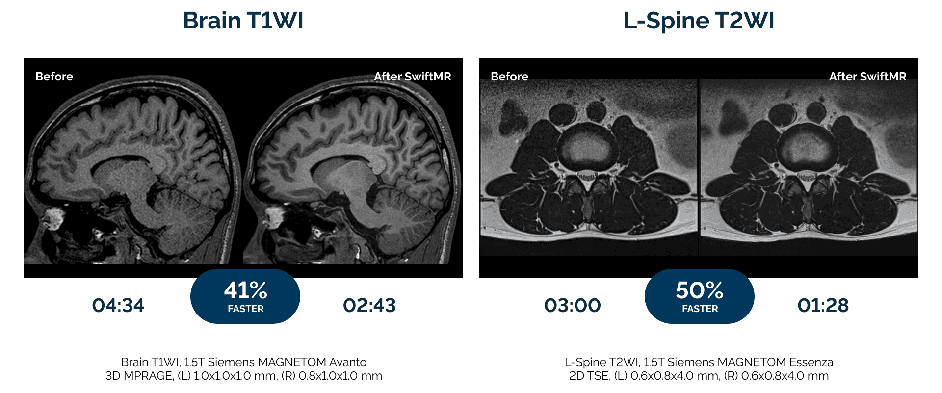
AIRS Medical built SwiftMR, AI-powered MRI reconstruction software, to meet today’s imaging challenges. Hospitals and clinics in over 35 countries use SwiftMR to:

  • Reduce scan times by up to 50% compared to standard protocols.
  • Deliver sharper images radiologists can trust.
  • Enhance the patient experience with shorter exams and fewer motion-related rescans.

SwiftMR is vendor-neutral, compatible with all MRI makes, models, and field strengths.

FDA-cleared, MDR-certified, and clinically validated, SwiftMR is trusted by over 300 imaging providers in the U.S. and over 1,000 globally, including:

These outcomes show that AI-powered MRI delivers tangible operational, clinical, and financial benefits across site types and geographies. 

Watch this video to learn more about SwiftMR.

The Takeaway

Radiology leaders are relying on SwiftMR to transform how they deliver care. From enterprise networks to single-scanner clinics, imaging teams are unlocking new levels of efficiency and patient care.

AI-Driven Lung Cancer Screening and Improving Patient Outcomes

AI is reshaping clinical decision-making, optimizing resource allocation, and enhancing both patient outcomes and experience in CT lung cancer screening. Radiology providers are successfully integrating new AI software tools into hospital operations – supporting diagnostic accuracy and improving patient outcomes.

At the center of this trend is Coreline Soft’s FDA-cleared AVIEW LCS Plus, a 3-in-1 solution capable of detecting lung nodules, quantifying emphysema, and analyzing coronary artery calcification – all from a single low-dose CT scan. 

  • AVIEW LCS Plus is in use at Temple Health, a nationally recognized institution in the U.S. Northeast, where it has allowed providers to streamline clinical workflows from detection to follow-up, delivering measurable improvements in care and ROI.

Coreline Soft will co-host a strategic webinar with the Temple Lung Center on August 1 at 1:30 PM ET, focused on AI-powered lung cancer screening and the evolving paradigm of early detection for chest diseases.

The webinar will offer firsthand insight into how Temple Health is drawing attention as a model for integrating AI beyond diagnosis – transforming it into a scalable, patient-centered care strategy.

The discussion will focus on two main areas…

  • Real-world outcomes: How AI improved diagnostic efficiency, early detection, and comorbidity detection.
  • A deep dive into the precision technology of the AVIEW LCS Plus platform.

AI like Coreline’s is not replacing clinical judgment, but reinforcing it, enhancing radiologists’ ability to detect, triage, and treat lung disease earlier and more efficiently, Criner believes. 

  • The webinar is open to pulmonologists, radiologists, cardiologists, respiratory-adjacent professionals, hospital stakeholders and administrators, and primary care providers across the U.S. and Canada. Interested participants can register for free in advance via the official registration link. 

The Takeaway

AI solutions like Coreline Soft’s AVIEW LCS Plus platform are having a real-world impact on healthcare providers as they roll out CT lung cancer screening programs. Sign up to learn more on August 1.

Molecular MRI Adds Certainty to Cancer Diagnosis

MRI has become an important tool in the detection, diagnosis, and treatment planning for many cancers, especially solid tumors. However, up until now, a lack of specificity has held back the full potential of MRI.  

While MRI is very good at identifying areas of interest, factors such as infection, benign tumors, post-traumatic areas, and inflammation can all increase vascularity and, therefore, enhancement of contrast and signal changes.  

  • As a result, MRI has a high rate of false positives – findings that may be flagged as something of concern but that are not necessarily malignant lesions.  

This lack of accuracy results in clinical care teams performing too many confirmatory biopsies, with most being benign.

Now a novel class of molecular imaging contrast agents developed by Imagion Biosystems brings a new level of specificity to MRI. 

  • The company’s MagSense imaging agents have the potential to improve the clinical utility of the large installed base of MRI systems across the globe through improved accuracy of interpretation, avoiding biopsies of benign lesions, driving earlier intervention and improving outcomes and quality of life.

Unlike gadolinium-based agents that non-specifically enhance tissue vascularity regardless of cause, MagSense imaging agents target receptors on cancer cells.  

  • By combining magnetic nanoparticles that have high susceptibility and r2 relaxivity with cancer-specific biomarkers, molecular MRI becomes possible.

Imagion’s superparamagnetic iron oxide nanoparticles are coated with a cancer-specific targeting moiety, such as an antibody or peptide.

  • The cancer biomarker molecule causes the particles to bind to target-specific cancer cells, if present. If the lesion in question is not the target cancer, the particles do not bind.

Where the imaging agent has become attached to the tissue, the nanoparticles produce an identifiable change in MRI signal. 

  • This signal is easily detected by radiological review and can be quantitatively assessed.

Imagion has developed cancer-specific contrast imaging agents for HER2 breast cancer, prostate cancer, and ovarian cancer, and the MagSense platform can be adapted for any type of cancer for which there is a targeting moiety.  

  • Imagion is now preparing to initiate a multisite phase 2 study in the U.S. in HER2+ breast cancer patients to optimize imaging parameters and compare MagSense imaging to the standard of care.  

The Takeaway

Molecular-specific imaging agents like the MagSense technology from Imagion Biosystems create the opportunity for molecular MRI to fundamentally change how radiologists detect and monitor cancers. 

The company is publicly traded (ASX:IBX) and is looking to expand its U.S. investor base as it advances through its clinical programs. To become involved as an investigator or investor or to learn more visit their website.

Integrated Solutions for Managing Incidental CAC Findings

The rising prominence of coronary artery calcium as a prognostic marker for heart disease has created an emerging challenge for radiologists: how should they manage incidental CAC findings discovered on routine CT exams? Fortunately, new industry collaborations are making it possible to deliver CAC reports to clinicians without disrupting workflow. 

Routine CT scans are revealing data beyond their original diagnostic intent.

  • AI solutions – such as AVIEW CAC from Coreline Soft – play a pivotal role in identifying risks for cardiovascular disease, osteoporosis, and metabolic disorders – all from a single scan.

AI allows one CT scan to assess lung, cardiovascular, and skeletal health, improving diagnosis and treatment planning.

One imaging services provider that has put AVIEW CAC into use is 3DR Labs, which has been actively integrating the solution into its nationwide clinical network.

  • The partnership enables 3DR Labs radiologists to generate consistent, high-quality CAC reports directly within PACS, while significantly reducing turnaround times.

3DR Labs is finding that AVIEW CAC optimizes workflow efficiency and significantly reduces the time required for CAC assessment. 

  • It also ensures that radiologic technologists can perform quick QA checks, enhancing consistency and reliability in the delivery of the report.

The latest generation of the FDA-cleared AVIEW CAC features an upgraded user interface and advanced batch-scoring functionality. 

  • 3DR Labs is now working to expand AI-driven insights into lung and neuroimaging through Coreline’s broader AVIEW platform (AVIEW ILA for interstitial lung abnormalities and AVIEW BAS for brain CT).

Beyond diagnostic imaging, this collaboration supports growing demands for cost-efficiency in healthcare. 

  • As U.S. insurers and government agencies recognize the ROI potential of early AI detection, platforms like AVIEW CAC offer scalable, high-performance solutions that lower costs and streamline care delivery.

3DR Labs has also highlighted Coreline Soft’s role as a founding partner in AI Labs, the company’s vendor-neutral platform to deliver the latest AI innovations to radiology workflows.

The Takeaway

New partnerships like the collaboration between Coreline Soft and 3DR Labs are advancing the future of AI in radiology – focusing on automation, early detection, and better patient outcomes through powerful, clinically validated technologies. Such partnerships not only reflect increasing adoption of AI in U.S. healthcare but set the stage for global transformation in diagnostic imaging.

Get every issue of The Imaging Wire, delivered right to your inbox.