MRI Accident Turns Deadly

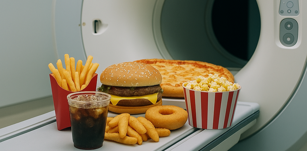
A tragic MRI accident in Long Island, New York, has turned deadly. A man who was pulled into a mobile MRI scanner by a heavy chain he was wearing died of his injuries. 

Keith McAllister was waiting outside a mobile MRI trailer operated by Nassau Open MRI on Long Island as his wife received a knee scan.

  • McAllister was wearing a weight-training chain around his neck that weighed some 20 pounds.

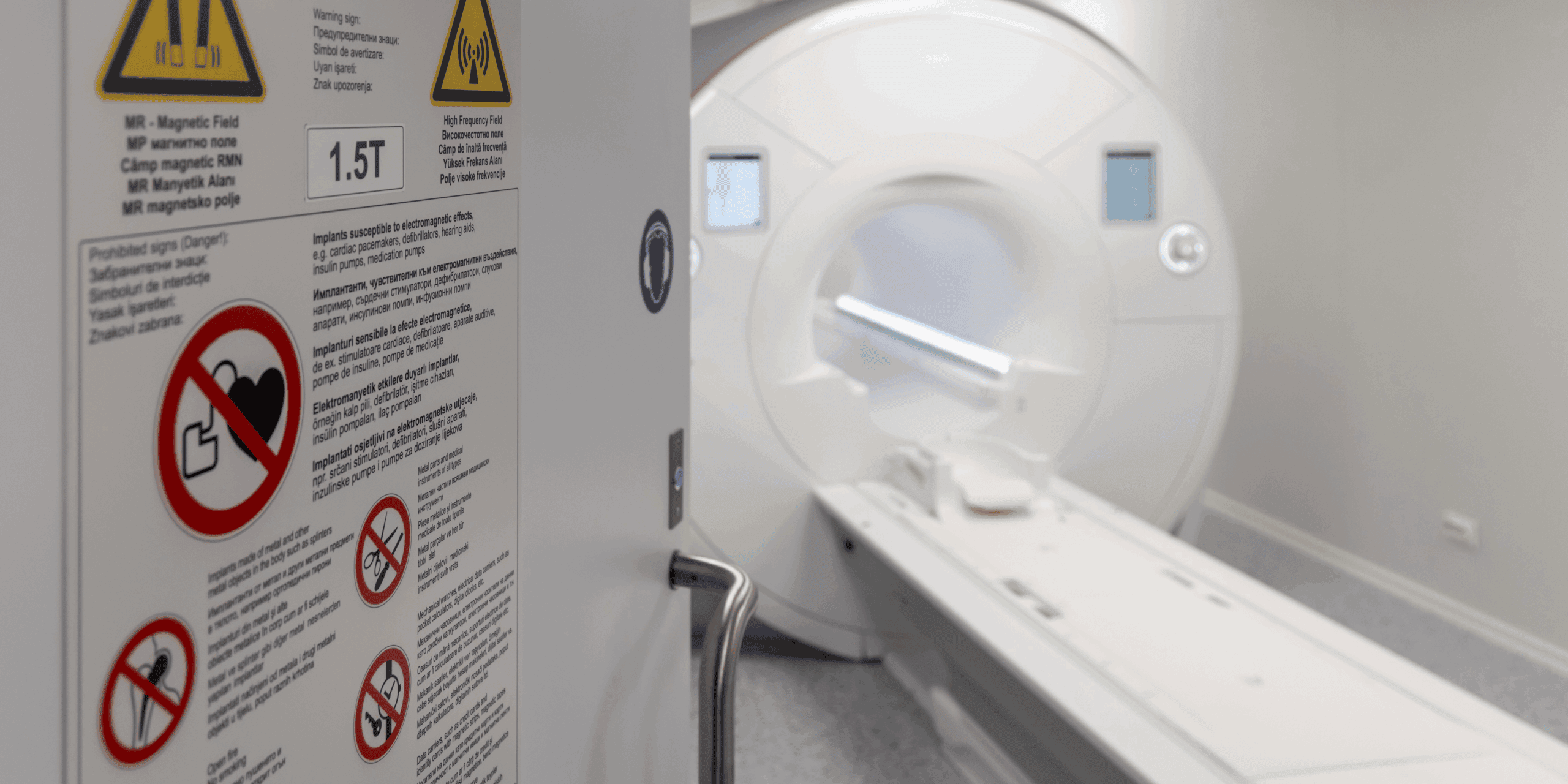
When he entered the trailer to help his wife get off the scanner table, the system’s powerful 1.5T magnetic field drew him against the magnet. It took staff an hour to free him.

Investigators are still looking into the details of the episode, but it underscores the shortcomings in how MRI safety is regulated in the U.S., where fatal MRI accidents are extremely rare but still do occur.  

  • That’s according to MRI safety expert Tobias Gilk, vice president at architectural firm Radiology Planning and founder of Gilk Radiology Consultants, who spoke to The Imaging Wire about the accident.

The U.S. has some of the most comprehensive and sophisticated guidelines on MRI safety, encapsulated in the ACR Manual on MR Safety.

  • What’s more, the radiology community including ACR, ISMRM, ASRT, and others are currently observing their annual MR Safety Week to promote safe MRI scanning – an event that started just a few days after McAllister died.

But despite the great leaps in knowledge about MRI safety, Gilk believes that keeping patients safe is complicated by the exponential growth in the modality’s complexity, while actual enforcement of safety standards is lacking. 

  • Many state health departments don’t even address MRI safety as they focus more aggressively on regulating ionizing imaging modalities like CT and X-ray, and healthcare certification bodies like the Joint Commission lack enforcement teeth.

Instead, MRI safety often becomes the responsibility of technologists who frequently must juggle multiple tasks as they manage both patients and scanner operations.

  • This can be particularly challenging in mobile MRI coaches, often staffed by a single MRI technologist where the only barrier between the outside world and the scanning environment is just a single – often unlocked – door. 

The Takeaway

The tragic death of Keith McAllister in a mobile MRI trailer shows that all the guidelines and safety events in the world won’t keep patients safe unless accompanied by stronger enforcement of the knowledge the radiology community already has. We can do better.

Prostate AI Improves Biparametric MRI

Researchers continue to hone in on the best way to use MRI for patients suspected of having prostate cancer, and AI is helping the effort. A new study in AJR shows that AI can improve the diagnostic accuracy and consistency of prostate MRI – while making it easier to perform.

Multiparametric MRI is the gold standard for prostate cancer imaging, but requires the use of three different MRI sequences as well as contrast administration, making it more complex and time-intensive to perform. 

  • On the other hand, biparametric MRI uses just two sequences – T2-weighted and diffusion-weighted imaging – and omits the contrast entirely, leading to shorter scan times and lower cost.

But what are you losing with bpMRI – and can AI help you get it back? Researchers addressed this question in the new study in which six radiologists interpreted bpMRI scans of 180 patients from multiple centers. 

  • Radiologists used a deep learning algorithm developed at the NIH to interpret bpMRI scans acquired on 3T scanners. The open-source algorithm generates binary prostate cancer prediction maps that are overlaid on T2-weighted images.

Researchers found that radiologists using the bpMRI AI algorithm to detect clinically significant prostate cancer had…

  • An increase in lesion-level positive predictive value (77% vs. 67%).
  • But lower lesion-level sensitivity (44% vs. 48%). 
  • And no statistically significant difference in patient-level AUC (0.82 vs. 0.83, p = 0.61).
  • While inter-reader agreement scores improved for lesion-level and patient-level PI-RADS scores and lesion size measurements. 

What to make of the numbers? The authors pointed out that the study design – in which AI was used as a first reader – may have reduced AI’s performance.

  • In real clinical practice, AI would most likely be used as a sort of clinical spell checker, with AI results overlaid on images that radiologists had already seen. 

The researchers said the results on improved positive predictive value and inter-reader agreement show that AI can improve the diagnostic accuracy and consistency of bpMRI for prostate cancer. 

The Takeaway

The new findings echo other research like the PI-CAI study highlighting the growing role of AI in prostate cancer detection. If validated with other studies, they show AI-assisted bpMRI could be ready to take on mpMRI for a broader role.

RP Builds AI Mosaic as Company’s IT Foundation

Radiology Partners announced a new initiative to guide the rollout of AI across its nationwide network of radiology practices. The company’s new MosaicOS will be the IT foundation that connects RP practices and supports clinical uses from AI-assisted reporting to report generation and even image management.

Radiology Partners has grown since its founding in 2012 to become the largest privately held provider of imaging services in the U.S. and a major force behind the consolidation of private-practice radiology groups.

  • RP has always maintained a heavy technology investment, and has been looking closely at the rise of AI in radiology.

That’s because the growth in imaging volume is so massive that clinicians will no longer be able to care for patients adequately without AI’s assistance, at least according to RP’s Associate Chief Medical Officer for Clinical AI Nina Kottler, MD.

RP laid the groundwork for MosaicOS in 2020 by first migrating its technology stack to a cloud-native infrastructure. 

  • This frees RP from reliance on on-premises legacy software and enables the company to push out updates that can be adopted quickly across its network.

RP’s Mosaic rollout includes the following components as the company…

  • Forms a new division, Mosaic Clinical Technologies, to oversee its AI activities.
  • Debuts MosaicOS, a cloud-native operating system that combines AI support with workflow and other IT tools.
  • Launches Mosaic Reporting, an automated structured reporting solution that combines ambient voice AI with large language model technology.
  • Develops Mosaic Drafting, a multimodal AI foundation model that pre-drafts X-ray reports that radiologists can review, edit, and sign. 

Mosaic Reporting is already in use at some RP sites, and the company is pursuing FDA clearance for broader use of Mosaic Drafting. More Mosaic applications are on the way.

  • Mosaic tools will be disseminated to RP centers using the cloud-native infrastructure, and MosaicOS will include image management functions that providers can choose to use in place of or alongside existing tools like viewers and archives. 

Kottler told The Imaging Wire that RP has de-emphasized individual pixel-based AI models in favor of foundation models that have broader application.

  • What’s more, RP CEO Rich Whitney said the company has chosen to develop AI technology internally rather than rely on outside vendors, as this gives it greater control over its own AI adoption.

The Takeaway

The launch of MosaicOS marks an exciting milestone not only for Radiology Partners but also for radiology in general that could address nagging concerns about clinical AI adoption on a broad scale. RP has not only the network but also the technology resources to make the rollout a success – the question is whether outside AI developers will share in the rewards.

Radiology AI Approvals Near 1k in New FDA Update

The FDA last week released the long-awaited update to its list of AI-enabled medical devices that have received marketing authorization. The closely watched list shows the number of AI-enabled radiology authorizations approaching the 1k mark.

The FDA has been tracking authorizations of AI-enabled devices going back to 1995, and the list gives industry watchers a feel for not only how quickly the agency is churning out reviews but also which medical specialties are generating the most approvals.

  • But the last time the FDA released an updated list was August 2024, and recent turmoil at the agency had some observers wondering if it would continue the tradition – as well as whether it could stay on pace for new approvals.

Those fears should be assuaged with the new release. The numbers indicate that through May 2025 the FDA has…

  • Granted authorization to 1.2k AI-enabled medical devices since it started tracking.
  • Approved 956 AI-enabled radiology products, or 77% of total medical authorizations.
  • Radiology’s share of overall authorizations from January to May 2025 ticked up to 78% (115/148), compared to 73% in the 2024 update, and 80% in all of 2023.
  • GE HealthCare remains the company with the most radiology AI authorizations, at 96 (including recent acquisitions like Caption Health and MIM Software), with Siemens Healthineers in second place at 80 (including Varian). 
  • Other notable mentions include Philips (42 including DiA Analysis), Canon (35), United Imaging (32), and Aidoc (30). 

In a significant regulatory development, the FDA said it was developing a plan to identify and tag medical devices that use foundation models, including large language models and multimodal architecture. 

  • The agency said the program would help healthcare providers and patients know when LLM-based functionality was included in a medical device (the FDA has yet to approve a medical device with LLM technology). 

In another interesting change, the FDA dropped “machine learning” from the title of its list, apparently with the idea that “AI” was sufficient as an umbrella term. 

The Takeaway

The FDA’s release of its AI approval list is a welcome return to past practices that should reassure agency watchers that recent turmoil isn’t affecting its basic operations. The LLM guidance suggests the agency may be changing its approach to the technology in favor of disclosure and transparency instead of more stringent regulation that could delay some LLM solutions from reaching the market.

AI-Driven Lung Cancer Screening and Improving Patient Outcomes

AI is reshaping clinical decision-making, optimizing resource allocation, and enhancing both patient outcomes and experience in CT lung cancer screening. Radiology providers are successfully integrating new AI software tools into hospital operations – supporting diagnostic accuracy and improving patient outcomes.

At the center of this trend is Coreline Soft’s FDA-cleared AVIEW LCS Plus, a 3-in-1 solution capable of detecting lung nodules, quantifying emphysema, and analyzing coronary artery calcification – all from a single low-dose CT scan. 

  • AVIEW LCS Plus is in use at Temple Health, a nationally recognized institution in the U.S. Northeast, where it has allowed providers to streamline clinical workflows from detection to follow-up, delivering measurable improvements in care and ROI.

Coreline Soft will co-host a strategic webinar with the Temple Lung Center on August 1 at 1:30 PM ET, focused on AI-powered lung cancer screening and the evolving paradigm of early detection for chest diseases.

The webinar will offer firsthand insight into how Temple Health is drawing attention as a model for integrating AI beyond diagnosis – transforming it into a scalable, patient-centered care strategy.

The discussion will focus on two main areas…

  • Real-world outcomes: How AI improved diagnostic efficiency, early detection, and comorbidity detection.
  • A deep dive into the precision technology of the AVIEW LCS Plus platform.

AI like Coreline’s is not replacing clinical judgment, but reinforcing it, enhancing radiologists’ ability to detect, triage, and treat lung disease earlier and more efficiently, Criner believes. 

  • The webinar is open to pulmonologists, radiologists, cardiologists, respiratory-adjacent professionals, hospital stakeholders and administrators, and primary care providers across the U.S. and Canada. Interested participants can register for free in advance via the official registration link. 

The Takeaway

AI solutions like Coreline Soft’s AVIEW LCS Plus platform are having a real-world impact on healthcare providers as they roll out CT lung cancer screening programs. Sign up to learn more on August 1.

CT Lung Screening Chats Pay Off

Patients who talked about CT lung cancer screening with their doctors were more likely to actually follow through on getting scanned. That’s according to a study this week in CHEST that offers support for shared decision making – a process that some screening proponents have criticized.

The U.S. continues to see disappointing compliance rates for CT lung cancer screening, over 10 years after the USPSTF recommended the exam.

  • Some lung screening proponents suggest that one barrier to screening is a CMS rule requiring a shared decision-making session between patients and doctors before the first scan is performed – a requirement that’s not in place for any of the other major cancer screening tests.

But the new study indicates that shared decision making could actually work to boost compliance. 

  • Researchers from the Harvey L. Neiman Health Policy Institute led by first author YoonKyung Chung, PhD, examined lung screening compliance rates for 22.6k people who had their first CT exam between 2016 and 2019.

Researchers looked at differences in annual follow-up lung screening rates between people who got shared decision-making sessions and those who didn’t, finding… 

  • Only 11% of study participants had a session before their first scan.
  • One year after the initial scan, those who participated in sessions were 27% more likely to get a follow-up exam.
  • Four years later, the compliance rate rose to 33%. 

If CMS requires shared decision-making sessions for reimbursement, why are they occurring so infrequently? 

  • The authors called this phenomenon “puzzling,” and suggested it’s because CMS is not enforcing the mandate through tools like claims denial. CMS could also boost utilization by providing higher reimbursement for the discussions.

The Takeaway

The new findings suggest that shared decision making should be viewed as an opportunity rather than a barrier to convincing patients of CT lung cancer screening’s value. The results track with other studies showing that a high-touch approach with tools like patient navigators can work.

Top 6 Radiology Trends from 2025’s First Half

The first half of 2025 has drawn to a close, and once again it was an eventful period for radiology. As we do every year, we’ve compiled a list of the top six stories – one for each month – to help recap what was important in medical imaging.

Consolidation in Imaging Services

Radiology’s imaging services segment continues to consolidate as smaller providers get gobbled up by larger players. One of the biggest consolidation moves happened in April when Radiology Partners agreed to take over radiology contracts held by Envision Healthcare. Private-practice radiology continues its slow decline, as documented by a study in May that found just 50% of U.S. radiologists in private practice.

DeepHealth Drives AI Consolidation

Imaging services isn’t the only radiology market that’s consolidating – the AI sector is also seeing heightened M&A activity as algorithm developers suffer from a decline in venture capital activity and slower clinical adoption. Many of the deals are being driven by RadNet’s DeepHealth subsidiary, which continues to acquire independent AI developers, such as See-Mode Technologies in ultrasound and its proposed deal for iCAD in mammography.

Radiology VC Funding Goes Boom – Then Bust

Radiology venture capital funding appeared to be gaining momentum in the first few weeks of 2025, as January saw six funding rounds, but the good times didn’t last as economic concerns slowed investment. Radiology startups may have to get used to a more competitive funding environment, although there was positive news in the spring with funding rounds from HOPPR, DESKi, RadAI, Yunu, Aeon, Heartflow, Chipiron, Brainomix, and Brainreader.  

When Will Getting Paid for AI Get Easier?

Reimbursement is one of the major stumbling blocks holding back wider adoption of clinical AI. Legislation that might grease the reimbursement skids was introduced into the U.S. Congress in April, but sources tell The Imaging Wire that it wasn’t included in the big budget bill that was just passed. In the meantime, AI developers and users will have to deal with a patchwork of AI reimbursement pathways.

Concerns Rise about CT Radiation Dose

Several studies were published in the first half of 2025 raising concerns about radiation dose from CT scans. First, researchers in April released a study that they claimed showed that all the CT scans performed in the U.S. in a single year would cause more than 100k cancers over the lives of the patients who got them. That was followed up with a paper in May linking greater CT use in European countries to a higher percentage of patients with five-year cumulative radiation dose of over 100 mSv. 

Can Imaging IT Tools Help Radiologists Manage Rising Volume?

Radiology’s rising scan volume and static workforce have IT developers furiously working on tools to bridge the gap. A March paper listed the half-dozen IT tools radiologists say they want (only one was AI), but another analysis threw cold water on the idea by predicting that AI would actually increase radiologists’ workload, not reduce it. Meanwhile, multiple studies are showing that for applications like breast screening, AI can reduce workload by as much as 41%.

The Takeaway

The midpoint of the year is a great time to take stock of radiology’s progress and the issues that have bubbled to the surface over the past six months. AI dominated radiology for the first half of 2025, and odds are the trend will continue in the back half of the year.

Get every issue of The Imaging Wire, delivered right to your inbox.

You might also like..

Select All

You're signed up!

It's great to have you as a reader. Check your inbox for a welcome email.

-- The Imaging Wire team

You're all set!