“The credo of ‘publish or perish’ has left a terrible battlefield in the papers on MRI.”
Radiology and MR professor, Dr. Peter Rinck, on the damage done from 30 years of often incorrect and ineffective MR research studies.
Imaging Wire Sponsors
- Carestream – Focused on delivering innovation that is life changing – for patients, customers, employees, communities and other stakeholders
- Focused Ultrasound Foundation – Accelerating the development and adoption of focused ultrasound
- Medmo – Helping underinsured Americans save on medical scans by connecting them to imaging providers with unfilled schedule time
- Nuance – AI and cloud-powered technology solutions to help radiologists stay focused, move quickly, and work smarter
- Pocus Systems – A new Point of Care Ultrasound startup, combining a team of POCUS veterans with next-generation genuine AI technology to disrupt the industry
- Qure.ai – Making healthcare more accessible by applying deep learning to radiology imaging
The Imaging Wire
Hologic Meets MARIA
Hologic signed a deal with Micrima to distribute the company’s MARIA radio-wave breast imaging system in Germany, Austria, and Switzerland. MARIA is highlighted for its radiation- and compression-free imaging, serving as an adjunct to mammography for locating cancers, particularly in younger women with dense breasts. Through the deal:
- Micrima Gets: A solid breast imaging distribution partner, as MARIA transitions from its product development phase into commercialization. This may not be limited to Europe’s DACH region, as Micrima was quick to reveal plans to launch “MARIA into further new territories.”
- Hologic Gets: An even more robust breast imaging portfolio, strengthening Hologic’s ability to be its clients’ only supplier and addressing the growing demand for dense breast imaging. In addition to R&D and acquisitions, Hologic has actively expanded its portfolio through distribution partnerships, including this deal with Micrima and its still-recent handheld ultrasound distribution alliance with Clarius.
DBT Doesn’t Need DM
New research in the American Journal of Roentgenology “strongly” suggests that digital breast tomosynthesis (DBT) could replace digital mammography (FFDM) as a standalone screening tool. This is far from the first study touting DBT’s advantages over FFDM, but this one may make the strongest case that DBT doesn’t need to be supported by FFDM. The researchers studied scans from 330 patients, finding that DBT:
- Performed better than FFDM among 29 of 31 readers, regardless of breast density
- Produced a “statistically significant” mean diagnostic accuracy improvement
- Achieved a higher subject-level AUC than FFDM (0.835 vs. 0.765)
- Posted a higher breast-level AUC than FFDM (0.861 vs. 0.818)
- Reduced noncancer recall rates per patient by 19%
- Detected more masses and architectural distortions than FFDM
- Detected invasive cancers with “significantly greater” accuracy than FFDM
Despite this solid evidence in favor of DBT (especially that final bullet), the researchers suggest that 2D overview images captured with FFDM will continue to be used along with DBT for the foreseeable future.
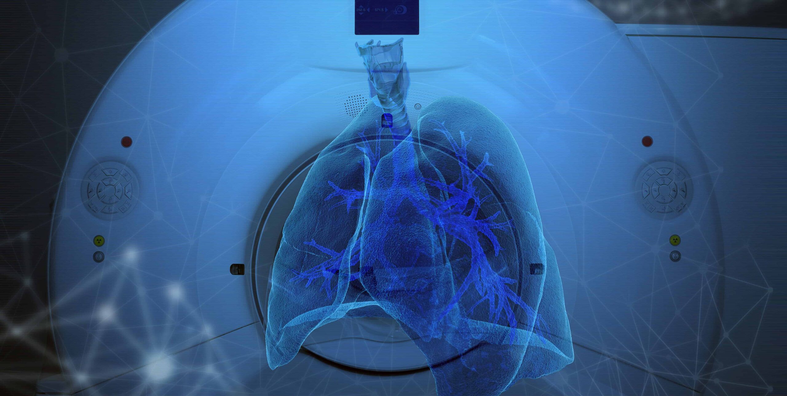
Questioning MR’s Progress
German radiology and MR professor, Dr. Peter Rinck, took to AuntMinnieEurope.com to criticize MRI’s slow and wasteful evolution. Dr. Rinck suggests that MRI functionality and applications have remained stable over the last 20 years, and despite advancements to MR hardware and software (and aesthetics), evolutions to the modality resulted in more unnecessary costs and procedures than clinical breakthroughs.
- Field Strength Overkill – Fifteen years after the first 3T MRIs became available, the majority of scans are still performed at 1.5T (2/3rds in U.S. and EU). However, even that may be overkill, given the sub-1.5T needs of most diagnostic situations.
- Ineffective Research – Dr. Rinck suggests that up to 95% of historic MR studies proved to be “simply wrong, senseless, and useless,” but still “influenced the use of MRI and medicine at large.” He doesn’t expect this trend to slow given the connection between research and career advancement.
- Irrelevant Innovation – The last few decades are full of MRI technology spin-offs that came and went (e.g. MR spectroscopy) or still haven’t proven to be effective (fMRI), leading to a cycle of euphoria and disillusionment that continues with each new development.
It feels better to end articles like this with some suggestions on the “good” things the industry can do with MRI technology. Although Dr. Rinck didn’t share any such advice, he might encourage field strength pragmatism, only performing and referencing valuable research, and focusing on value-based innovations.
Imaging Wire Q&A: Qure.ai’s Stroke Solution
In the first-ever Imaging Wire Q&A, we sat down with Qure.ai’s co-founder and Head of R&D, Dr. Pooja Rao, to discuss current challenges with treating stroke and head trauma and how AI solutions, such as Qure.ai’s qER product, stand to improve clinical outcomes. Here are some of the big take-aways:
- Motivation – Stroke is one of the leading causes of death and long-term disability worldwide and over 2.5 million people suffer head injuries annually in the U.S. alone. Imaging plays a key role in diagnosing both stroke and head trauma, while AI has the potential to speed up diagnosis and get treatment to the patients who need it most.
- AI Research – Standalone studies show that AI works well and is safe and effective enough to be used in clinical practice, and it sounds like regulatory bodies agree, given the recent clearance of AI products to triage critical scans and assist radiologists. Qure.ai’s own study showed that qER accurately detects not only bleeds but also other critical head CT scan abnormalities.
- Clinical Use AI – Early evidence shows that AI works just as well in clinical setting as it does in the lab, proving that it generalizes well, speeds treatment times, and improves radiologist efficiency. There has also been particularly strong early adoption among community hospitals and remotely located healthcare providers without reliable access to radiologists.
- Patient Benefits – For patients, a lot of the benefit of AI is access – just having access to rapid, accurate diagnosis and treatment, and not having to wait hours in the ER.
- Radiologist Benefits – For radiologists, the benefits of AI differ based on the setting in which they operate. Busy urban practices or teleradiology setups benefit the most from having critical cases automatically flagged for review. In places where radiologist coverage is sparse, radiologists and other clinicians find mobile phone alerts with non-diagnostic preview images particularly useful.
- The Next Frontier – Everyone wants algorithms that can be superhuman and see abnormalities that radiologists can’t, but there are easier problems to solve first. Two of these problems are incorporating clinical context and predicting long-term outcomes, which Qure.ai is working to build into its solutions.
The Wire
- New research from Johns Hopkins Hospital finds that primary care providers have increased their use of imaging for lower back pain, noting that much of these scans may have been unnecessary, ineffective, and likely to led to incidental findings. The study looked at 627,000 patients who visited PCPs with lower back pain, finding that the rate of imaging orders increased from 11% in 2011 to 14% in 2016, with 96% of these studies performed with X-ray (3% MRI, 2% CT).
- Frost & Sullivan reported that the global ultrasound market reached $6.12 billion and 115,592 units in 2018, led by the cart-based segment (73.4% revenue share, 60.4% unit share). The firm forecasts that emerging ultrasound segments (e.g. musculoskeletal, anesthesiology, and endocrinology) will become more established by 2023, while AI applications will be increasingly common for premium ultrasound applications (e.g. detecting breast cancer, fetal image assessment, cardiology).
- Denmark’s breast cancer screening scandal took a turn for the worse when it was revealed that up to 2,000 higher-risk women in the Jutland and Funen regions were only screened with mammography, despite a government guideline that their screenings should also include ultrasound and a doctor’s exam. The screening scandal was previously believed to be limited to a hospital in Denmark’s Zealand region and the new screening lapses are creating concerns that other regions might have similar issues.
- Reaction Data’s latest survey explored healthcare execs’ perspectives on cloud-based solutions (n=242), with a sizable 70% stating that the industry is headed towards cloud adoption, 14% believing cloud solutions are intriguing but not ready for prime time, and 10% stating that cloud solutions are over-hyped. Cloud solutions’ greatest advantages over on-premise solutions included lower support needs (~27 responses), moving from capex to opex (~24), and ease of deployment (~19), while the most prominent evaluation criteria include data security (29%), performance (26%), and financial impact (21%).
- Fujifilm Europe announced the upcoming launch of its new Mobile Mammography Unit, equipping a trailer with its AMULET Innovality DBT system. The Mobile Mammography Unit will primarily support providers while they are transitioning to a new mammography room (minimizing disruption and allowing treatment), but will also support procurement decisions by bringing the systems to the site of providers who are considering the AMULET Innovality DBT.
- Canon Medical Systems announced the Japan launch of the Aplio a Verifia ultrasound system, which has a value proposition that focuses on sonographer comfort, due to its height-adjustment features. The Aplio a Verifia announcement also touted its Protocol Assistant function (guides different procedures), 21.5-inch monitor (like Canon’s higher-end models), and its ability to image internal organs and blood flow.
- A recent IHS Markit report forecasts slow growth for the 3D mobile C-arm market. The report listed a number of challenges for 3D mobile C-arms including their focus on spinal and orthopaedic interoperative imaging (no vascular procedures), the dominance of Medtronic’s O-arm and hybrid operating rooms in the U.S., their high costs (4x higher than a 2D mobile C-arm), long processing and acquisition times, and a lack of professionals trained to interpret 3D images. These systems do have some advantages including supporting ongoing interventions, their wider adoption in Germany and China, and strong navigation capabilities, but these aren’t enough to drive significant growth.
- Avante Ultrasound took another step towards its goal of becoming a nationwide ultrasound system, service, and parts provider, unifying the operations of its Ultra Solutions (West coast – acquired in October) and Global Medical Imaging (East coast) operations under a single leadership team. The ultrasound unit’s integration also contributes to Avante Health Solutions overall strategy to become a major US and international player in medical device, service, and refurbishing (across modalities).
- Duke University researchers found that clinicians may be able to differentiate ductal carcinoma in situ (DCIS) from benign breast disease and reduce false-positive biopsy rates by measuring the growth of calcifications. The study used 74 DCIS calcifications and 148 benign calcifications, finding that the DCIS calcifications were larger (10mm vs. 6mm median) and grew at a much greater annual rate (96% vs. 68%) than benign breast calcifications.
- Direct-to-consumer imaging company, Ezra, launched its full-body MRI cancer screening program, expanding upon the prostate cancer screening program it launched in early 2019. Ezra’s subscription-based “members” pay $1,950 for full-body scans that can detect up to 11 cancers in men and 13 in women (single region scans are $675) at eight different New York City area facilities (more regions later in 2019), with plans to eventually support its below-market rates through AI efficiencies.
- Research from UCSF finds that radiologists are valuable collaborators with radiation oncologists, beyond tumor board settings. Looking at 122 cases presented at multidisciplinary thoracic tumor boards (TTB) and 45 cases at diagnostic radiology/radiation oncology rounds (DR/ROR), imaging-related discussions with a radiologist in DR/ROR settings led to more changes in radiation oncology management (68.9% DR/ROR vs. 31.1% TTB), more changes in radiation therapy timing (46.7% vs. 31.1%), and more changes to target volume (35.6% vs. <1%). DR/ROR imaging discussions also increased radiation oncologists’ confidence in 95.6% of cases and resulted in at least “moderate” change in radiation oncology management in over half of all DR/ROR discussions.
The Resource Wire
– This is sponsored content
- In this Carestream video, orthopaedic surgeon Dr. Bryan Den Hartog presents clinical images illustrating traditional CT vs. extremity CT imaging and discusses how the image resolution in the OnSight 3D Extremity System helps in his practice.
- Focused ultrasound system developer, Profound Medical, announced the final results from its TULSA-PRO prostate cancer ablation clinical trial, achieving a 94.9% median prostate-specific antigen (PSA) reduction and reducing PSA in 95.7% of all patients treated.
- This Nuance Healthcare Diagnostics Q&A details radiology AI’s “last mile” adoption challenge and outlines ways that Nuance and radiologists are overcoming these challenges.
- Qure.ai’s Rohit Ghosh takes the Tedx stage to discuss using artificial intelligence to tackle India’s TB problem.
- POCUS Systems’ forthcoming ultrasounds will combine ease of use, durability, and reliability, allowing clinicians to focus on their patients.
- A new study in JACR revealed that the rise of high-deductible health plans has led to greater patient concerns over imaging costs than ever before, while patient cost comparisons often leads to “confusion, misinformation, and opaqueness.” These are the exact patients who can be helped by the Medmo platform, which connects high-deductible patients with radiology centers, ensuring the best value for patients and a profitable revenue stream for imaging centers.