|
Learning Curve in DBT Screening | Radiology’s Spring Rebound
May 8, 2023
|
|
|

|
|
Together with
|

|
|
|
“Radiology has a history of innovation. It has defined our profession in many ways and I’m proud of that. “
|
|
James Backstrom, MD, of Armstrong County Memorial Hospital in the latest edition of The Imaging Wire Show on MRI efficiency.
|
|

|
|
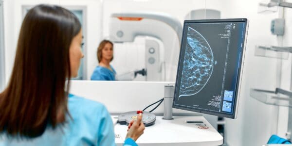
Digital breast tomosynthesis continues to evolve. First introduced initially as a problem-solving tool in breast imaging, DBT is becoming the workhorse modality for breast screening as well.
But DBT still requires some adjustment when used for screening. In a study of nearly 15k women in European Radiology, Swedish researchers describe how the false-positive recall rate for DBT cancer screening started higher but then fell over time as radiologists got used to the appearance of lesions on DBT exams.
The Malmö Breast Tomosynthesis Screening Trial was set up to compare one-view DBT to two-view digital mammography for breast screening. Unlike some DBT screening trials, the study did not use synthesized 2D DBT images. DBT images were acquired 2010-2015 with Siemens Healthineers’ Mammomat Inspiration system.
Findings in the study included:
- DBT had a sharply higher false-positive recall rate in year 1 of the study compared to DM (2.6% vs. 0.5%)
- DBT’s recall rate fell over the five-year course of the study, stabilizing at 1.5%
- Recall rates for DM varied between 0.5% and 1% over five years
- Most of the DBT recalls (37.3%) were for stellate lesions, in which spicules radiate out from a central point or mass. With DM, only 24.0% of recalls were for stellate lesions
- The number of stellate distortions being recalled with DBT declined over time, a trend the authors attributed to a learning curve in reading DBT images
The authors said that the DBT false-positive recall rate in their study was “in general low” compared to other European trials. They claimed that MBTST is among the first studies to analyze recall rates by lesion appearance, an important point because radiologists may see a different distribution of lesion types on screening DBT compared to what they’re used to with DM.
The Takeaway
The Malmö Breast Tomosynthesis Screening Trial was one of the first to investigate DBT for breast screening, and previous MBTST research showed that DBT can also reduce interval cancers, which occur between screening rounds.
The new findings offer further support for DBT breast screening and give hope that whatever shortcomings the technology might have early on in a screening role can be addressed through training and experience. It also confirms recent research indicating that DBT has become the new gold standard for breast screening.
|


|
|
Improve Your Cardiovascular Image Data Management
Every 34 seconds, a patient dies of cardiovascular disease. Find out how you can better manage cardiovascular image data and improve patient care with syngo Dynamics from Siemens Healthineers.
|
|
Creating a Modern Imaging Network in Costa Rica
SOIN Soluciones Integrales of Costa Rica turned to Merge enterprise imaging solutions from Merative when it wanted to modernize the imaging environments of 50 hospitals across the country. Download this PDF white paper to find out how they did it.
|
|
- TTG Buys Digirad: Digital gamma camera pioneer Digirad Health has been acquired by TTG Imaging Solutions, a Pittsburgh-based provider of molecular imaging equipment and radiopharmaceuticals. Digirad was one of the first companies to sell a solid-state digital SPECT camera and has focused on cardiac imaging. TTG has grown by acquisition of late, buying Acceletronics and RadPath in late 2022 and Medical Imaging Technologies in 2021. Digirad expands TTG’s presence on the West Coast and in cardiology and radiation oncology.
- Pandemic Prompts Payment Shift: The COVID-19 pandemic caused a major change in industry payments to radiologists, say researchers in JACR, with more money circulating to fewer radiologists. From 2019 to 2020, the number of radiologists getting paid fell 32% (15,170 vs. 10,323), but total payment value grew 37% ($68.2M vs. $93.6M) before falling back to $64.9M in 2021. Authors say the pandemic led to fewer gifts and speaking fees and more royalty/ownership payments.
- AR Tool Helps ‘See’ Radiation: What if you could “see” radiation levels in the environment around you? An augmented reality tool developed by Oak Ridge National Laboratory and licensed to Teletrix leverages gaming technology to divide physical space into volumes based on ionizing radiation dose. Users wearing AR headsets can see dose levels as color-coded gradients overlaid on physical space. The technology is focused on workplace safety like in the nuclear industry, but it could have medical applications such as for interventional radiology.
- AI Aids Portable Breast Ultrasound: An AI algorithm analyzing breast ultrasound images performed well identifying cancer regardless of whether images were acquired on low-cost portable units (96% sensitivity) or high-end cart machines (98% sensitivity). In a study from Mexico published in Radiology, Koios Medical’s Koios DS algorithm analyzed 1,216 breast masses found in 478 women from 2017 to 2021. The findings indicate that AI and portable breast ultrasound can be used as triage tools in low-resource settings.
- 10 Radiology Use Cases for ChatGPT: Ten possible use cases for ChatGPT in radiology are outlined in a fascinating Twitter thread by Woojin Kim, MD, a radiologist at the Palo Alto VA Medical Center and CMIO at Rad AI. Topping the list is summarizing patient records to supplement clinical indications; other top uses are summarizing priors and generating structured reports with minimal input. Kim sees radiology reporting as “ripe for disruption” as the specialty works to put ChatGPT to use.
- FAPI-PET’s Superiority: PET/CT imaging with a 68Ga-FAPI radiotracer was superior to FDG in detecting multiple types of cancer, says a new study in Journal of Nuclear Medicine. Researchers from China and Singapore tested 68Ga-FAPI-RGD in 22 patients with cancer types ranging from lung to ovarian. The radiotracer showed better tumor uptake (SUVmax = 18.0 vs. 9.1) and tumor-to-background ratio (15.2 vs. 5.5) compared to FDG, thanks to its affinity for the integrin αvβ3 tumor receptor.
- Annalise Opens India Office: AI developer Annalise.ai is continuing its global expansion by opening an office in India, Annalise India Centre in Chennai, to focus on developing new AI products. The company notes that India is one of the fastest-growing medical device markets, and is home to a large and diverse talent pool that can be tapped to develop new AI applications to complement its Annalise Enterprise CTB for CT brain and Annalise Enterprise CXR for chest X-ray applications.
- Practice Sues after Cyberattack: A North Carolina radiology group is suing its insurance broker after it suffered a ransomware attack just weeks after the group’s cyberattack coverage expired. Raleigh Radiology Associates charges that the broker failed to renew its $10M cyberattack protection policies just before the attack in February 2022. Raleigh had no coverage and was left “on its own” to respond to the attack, and is claiming over $1M in damages. The suit illustrates the challenges cybersecurity presents to small practices.
- ScreenPoint Lands Mid-Atlantic Practice: ScreenPoint Medical will be installing its Transpara software at Fairfax Radiology, which provides imaging services in the Washington, DC region. Fairfax is the largest radiology practice in the DC area, with almost 100 subspecialized radiologists and a breast imaging division with 18 dedicated breast imagers. Transpara is cleared for use with 2D and 3D mammography cases, providing a “second set of eyes” to detect breast cancer earlier and reduce recall rates.
- NIH Grant to Fund AI for Cardiac CT: The NIH apparently sees potential in cardiac CT AI, providing a $6.2M grant to support a Case Western and University Hospitals Cleveland Medical Center project that will develop AI tools to analyze cardiac CT images and predict cardiovascular risks. The project will leverage cardiac CT exams from UH’s CLARIFY Registry, which began in 2017 when the medical center began offering high-risk community members free CT calcium score exams, although the team will study all aspects of these images.
- New C-Arm Launched: Philips has launched Zenition 10, a new mobile C-arm for performing image-guided interventional procedures. The C-arm employs a flat-panel digital detector and is positioned as a cost-effective option for minimally invasive procedures in orthopedics, trauma, and other surgical areas. Zenition 10 includes a DSA capability, a low-dose pediatric mode, and image processing algorithms to reduce metal implant artifacts. The new C-arm complements Zenition 50 and Zenition 70 in the company’s C-arm portfolio.
|
|
Steps to Monetizing Your Medical Image Data
Looking to monetize your medical imaging data by sharing it with researchers or vendors? You’ll need to take care of a few things first, like de-identifying records and removing protected health information. Find out how to do it in this white paper from Enlitic.
|
|
Blackford’s AI Value Matrix
Working out your AI business case? Check out this helpful Blackford Analysis post detailing how to create your AI Value Matrix based on your organizational objectives and value indicators.
|
|
- See what UC Davis has to say about United Imaging’s EXPLORER Total Body Scanner, the first whole-body PET. EXPLORER has an effective sensitivity for total-body PET that’s 40-fold higher than current commercial scanners.
- If you’re at SIIM 2023, be sure to attend an InformaticsTECH Talk on Wednesday, June 14 on the Power of the Platform for Imaging AI, by Aaron Sullivan, Head of Business Development for Bayer Digital Solutions.
- Radiology faces numerous challenges to more efficient workflow, from the siloed nature of healthcare enterprises to mundane tasks that are ripe for automation. In this Imaging Wire Show, we talked to Dr. Matthew Lungren and Calum Cunningham of Nuance Communications.
- What’s behind healthcare’s shift from legacy PACS to cloud-based enterprise imaging? We talked to Brad Levin of Visage Imaging at HIMSS 2023, and he explains the change and the benefits of Visage’s Visage 7 | CloudPACS solution.
- “It completely changes the way we think about MRI imaging.” Take a look at this video interview with Mass General’s Chief of Neurocritical Care to see how clinicians can use Hyperfine’s Swoop Portable MRI to eliminate care disruptions in the ICU by keeping critically ill patients in the unit throughout the neuroimaging process.
- Radiologist recruiting and retention is more important than ever in today’s hyper-competitive radiologist job market. Learn from industry experts and practice leaders in this on-demand Medality webinar as they reveal how to overcome hiring challenges, keep your team engaged, and provide opportunities for growth.
- What is the Signa Experience? It’s a suite of diagnostic solutions for every step of a patient’s MRI pathway, from the newly launched Signa One user interface to AIR Recon DL data reconstruction for noise removal to light and flexible AIR Coils. Learn more from GE HealthCare.
- Annalise.ai’s Annalise CXR solution detects up to 124 findings in a single chest X-ray. See how it detects such a wide range of abnormalities using these demo studies… or upload your own CXR images.
- Faced with rising scan volumes and many elderly patients, Lake Medical Imaging implemented Subtle Medical’s Subtle MR efficiency solution across its eight MR scanners, allowing it to scan 40 additional patients per day while maintaining quality of care.
- Imaging AI is evolving fast, but radiology leaders’ expectations for their AI technologies might be evolving even faster. In this Imaging Wire Show with Dr. Charlene Liew of SingHealth and Dr. Nina Kottler of Radiology Partners, we explore radiology leaders’ current and future expectations for AI, and the central role platforms play in their AI roadmaps.
- Us2.ai just published what might be the most comprehensive paper we’ve seen on AI echo, detailing the benefits of AI-automated echocardiography, the global need for more scalable and flexible CVD assessments, and how its technology is fit for the future.
- Medical image sharing technology from Intelerad made it possible to find faster organ matches at Gift of Life Michigan. Medical images of organ viability are now available with other clinical data, speeding the match between donor and recipient.
|
|
|
Share The Imaging Wire
|
|
Spread the news & help us grow ⚡
|
|
Refer colleagues with your unique link and earn rewards.
|

|
|
|
|
Or copy and share your custom referral link: *|SHAREURL|*
|
|
You currently have *|REFERRALS|* referrals.
|
|
|
|
|