|
Pancreatic Cancer Radiomics | Finding a CT Alliance
July 17, 2022
|
|
|

|
|
Together with
|

|
|
|
“Maybe one day it’ll be the Google Maps of imaging, when we go where it tells us. Now we’re still: ‘no, I’ll take my own route.’”
|
|
OHSU cardiologist and SCCT 2022 panelist Maros Ferencik, describing cardiac imagers’ current and future reliance on AI.
|
|
|
ICYMI: Our third newsletter – Cardiac Wire – launched early last week. If you’re interested in cardiology and want the most important news curated and written with the typical “Wire” style, subscribe to get Cardiac Wire.
|
|

|
|
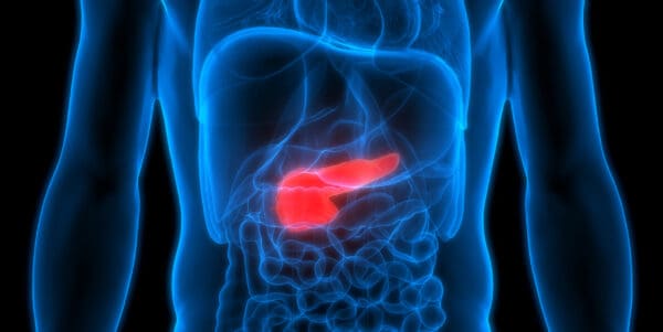
Mayo Clinic researchers added to the growing field of evidence suggesting that CT radiomics can be used to detect signs of pancreatic ductal adenocarcinoma (PDAC) well before they are visible to radiologists, potentially allowing much earlier and more effective surgical interventions.
The researchers first extracted pancreatic cancer’s radiomics features using pre-diagnostic CTs from 155 patients who were later diagnosed with PDAC and 265 CTs from healthy patients. The pre-diagnostic CTs were performed for unrelated reasons a median of 398 days before cancer diagnosis.
They then trained and tested four different radiomics-based machine learning models using the same internal dataset (training: 292 CTs; testing: 128 CTs), with the top model identifying future pancreatic cancer patients with promising results:
- AUC – 0.98
- Accuracy – 92.2%
- Sensitivity – 95.5%
- Specificity – 90.3%
Interestingly, the same ML model had even better specificity in follow-up tests using an independent internal dataset (n= 176; 92.6%) and an external NIH dataset (n= 80; 96.2%).
Mayo Clinic’s ML radiomics approach also significantly outperformed two radiologists, who achieved “only fair” inter-reader agreement (Cohen’s kappa 0.3) and produced far lower AUCs (rads’ 0.66 vs. ML’s 0.95 – 0.98). That’s understandable, given that these early pancreatic cancer “imaging signatures” aren’t visible to humans.
The Takeaway
Although radiomics-based pancreatic cancer detection is still immature, this and other recent studies certainly support its potential to detect early-stage pancreatic cancer while it’s treatable.
That evidence should grow even more conclusive in the future, noting that members of this same Mayo Clinic team are operating a 12,500-patient prospective/randomized trial exploring CT-based pancreatic cancer screening.
|


|
|
DASA and CARPL.ai’s Pediatric AI Evaluation
When Sao Paolo’s Diagnosticos da America SA (DASA, the world’s 4th largest diagnostics company) set out to evaluate Qure.ai’s QXR solution for their pediatric chest X-ray workflows, they leveraged CARPL.ai’s platform to streamline their evaluation. See how it worked here.
|
|
Three Questions that May Change your Enterprise Imaging Strategy
To solve the challenges of enterprise imaging, you’ll need a strategy that addresses today’s needs and future challenges. Answer the questions to see if you’re prepared to formulate a more effective healthcare IT plan.
|
|
- Oxipit & contextflow’s CT Alliance: Oxipit and contextflow announced plans to combine Oxipit’s ChestEye Quality AI solution and contextflow’s SEARCH Lung CT software, creating a combined solution to detect missed findings in chest CTs. ChestEye Quality has traditionally been used as a CXR double reader, notifying radiologists of potential missed findings when it identifies a mismatch between CXR images and radiologist reports. Integrating contextflow’s CT image analysis capabilities will allow ChestEye Quality to similarly identify and flag missed nodules in chest CTs.
- CAD-RADS 2.0: The major cardiology and radiology societies released their updated CAD-RADS 2.0 consensus document, expanding on the 2016 coronary CTA reporting guidelines. CAD-RADS 2.0 brings a critical update emphasizing that plaque burden should be estimated whenever present through visual assessment, segment involvement score, coronary artery calcium evaluation, or total plaque burden quantification. Other updates include new modifiers, including assessment of FFR-CT or myocardial CT perfusion when performed.
- GLEAMER & Fujifilm’s X-Ray AI Integration: GLEAMER and Fujifilm launched a partnership that will allow GLEAMER’s BoneView AI fracture detection solution to be integrated into any Fujifilm X-ray system that’s equipped with the OEM’s EX-Mobile image processing box. The combined solution will bring AI fracture detection to the point-of-care, allowing imaging teams to triage and rout patients with fractures within 30 seconds. Fujifilm has been actively expanding its AI-embedded X-ray partnerships, following CXR AI integration alliances with Qure.ai and Annalise.ai.
- CTPA Pulmonary Hypertension Detection: A recent Radiology study suggests that CTPA-based assessments of pulmonary vessel volumes could be used to identify patients with pulmonary hypertension (PH). Researchers performed quantitative CTPA assessments on 1,823 patients (1,593 w/ PH, 230 controls), finding that patients with pulmonary arterial hypertension and PH associated with left-sided heart disease also had lower average pulmonary vessel volumes. However, the PH patients and healthy controls had similar peel vessel volumes.
- Intelerad’s TA Investment: Intelerad announced an investment from private equity firm TA Associates, joining its existing investors (Hg & ST6), and setting Intelerad up for “the next phase of its growth journey.” Intelerad has been one of imaging informatics’ most-aggressive acquirers in recent years, enhancing its capabilities across the enterprise (cardiology, OB/GYN), use cases (imaging sharing, cloud), and global regions (UK). This new funding will be used to support both organic growth and allow Intelerad to pursue “strategic growth opportunities.”
- Rock Health’s H1 Slowdown: Rock Health confirmed that the first half of 2022 brought a significant slowdown in digital health venture funding, falling to $10.3B. Digital health startups are now projected to raise $21B through all of 2022, which is far below 2021’s inflated levels but still well above 2020’s total funding ($29.1B & $14.7B). Although a relatively small portion of H1’s funding went to imaging companies, three of the six most-funded clinical indications involve imaging (oncology #2, cardiovascular #3, neurology #6).
- Affidea Expands: European imaging center giant Affidea announced a series of center acquisitions in Spain (3), Portugal (2), and Romania (2), increasing its presence to 327 imaging centers in 15 countries. Affidea has already expanded into 6 new countries this year, helped by the support of its new parent company, GBL.
- ACTA AI Contrast Reduction: A new European Radiology study detailed an AI reconstruction approach that could allow aortic CT angiography using ultra-low contrast volumes (30% of normal dose). The researchers trained an Au-CycleGAN algorithm using ultra-low-dose and low-dose ACTA exams from 100 patients with suspected aortic disease, and then used it to reconstruct 50 ultra-low-dose ACTAs. The AI-reconstructed ultra-low-dose ACTAs scored better image quality than the unenhanced images from both groups (except for image noise), while producing images with no significant diagnostic differences.
- Siemens Integrates SubtleMR: Subtle Medical’s SubtleMR DLIR software is now officially integrated into Siemens Healthineers’ OpenRecon interface, making SubtleMR available with all newer Siemens MRI scanners once OpenRecon gains FDA clearance (targeting H2 2022). OpenRecon enables the integration of third-party image reconstruction and post-processing solutions (like SubtleMR), eliminating additional export steps and making results directly available on the scanner console.
- NL’s AI Adoption: An ECR presentation covered by Aunt Minnie detailed Dutch hospitals’ increasing imaging AI adoption. The survey of 29 healthcare organizations in the Netherlands (out of 69 contacted), revealed that 24 are using AI in clinical practice (vs. 23 of 37 respondents in 2021, 14 of 44 in 2020). These hospitals are using 46 unique imaging AI products (vs. 39 in 2021, 8 in 2020) and have 74 total implementations (vs. 49 in 2021, 19 in 2020).
- Shimadzu’s New MobileDaRt: Shimadzu Medical Systems USA officially launched its latest portable X-ray system, the MobileDaRt Evolution MX8 V. The new X-ray system’s features focus on maneuverability (motorized wheels, support column folds for visibility), its built-in 19in monitor, and its new “V type” X-ray panel (joins three other FPD panel sizes).
|
|
- Hyperfine’s MRI accessibility advantage doesn’t stop at image acquisition. See how Hyperfine’s BrainInsight AI tools give clinicians the quantitative biomarker information needed to streamline patient care and reduce the burden of manual brain MRI measurements.
- Wondering when someone will finally develop AI to address hanging protocol issues? The Enlitic Curie platform uses a standard imaging lexicon and a sophisticated algorithm to standardize DICOM metadata, making hanging protocols and automatic comparisons more consistent, and allowing radiologists to focus on image interpretation.
- See how Novarad’s CryptoChart solution allowed Central Ohio Primary Care (COPC, 70 practices, 400 physicians) to make the transition to digital imaging sharing in this Healthcare IT News case study.
- We hear a lot about AI being the next big thing or being immature and overhyped. This set of Blackford Analysis editorials reviews the challenges that are still holding back imaging AI, and the areas that AI is delivering genuine clinical benefits.
- With radiation dose management now largely considered best practice, this Bayer white paper details the top five benefits of adopting contrast dose management.
|
|
|
|
|